Figure 7. Effect of FBS on RPC expression
of markers as shown by immunocytochemistry. RPCs were grown in the
absence (A, C, E, G, I, K)
or presence (B, D, F, H, J, L)
of FBS for seven days, fixed, and immunolabeled with antibodies against
nestin (A, B), Ki-67
(C, D), β3-tubulin (E, F), GFAP (G,
H), rhodopsin (I, J); and PKC-α (K,
L). Final cell densities in chamber slides were dependent upon
treatment conditions. Cell nuclei were counterstained with DAPI. Scale
bars represent 100 μm.
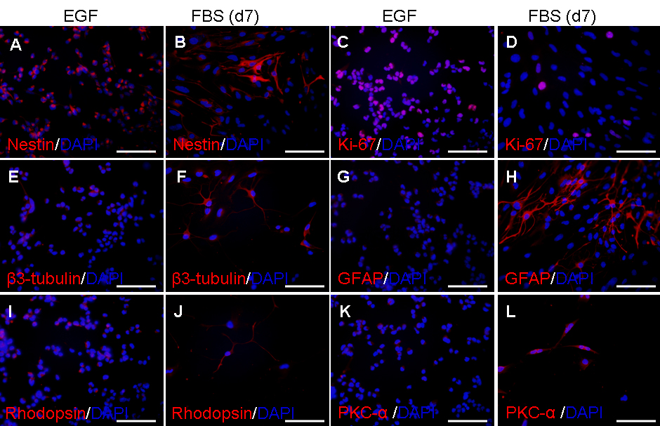
 Figure 7 of Gu, Mol Vis 2009; 15:2111-2122.