Figure 5. Cells isolated from each of
three scaffolds were serially propagated for three passages. This
allowed us to obtain information about the residual clonogenic
potential of the cells grown onto the scaffolds and to evaluate the
effects of the matrix on the preservation of stemness and induction of
differentiation pathways. [HKL + 3T3/J2] = HKLs with 3T3-J2 feeder
layer; [HKL - 3T3/J2] = HKLs without 3T3-J2 feeder layer. No difference
in the number of clonogenic cells (A) or percentage of aborted
colonies (aborted colonies/total colonies ratio; B) was
observed when cells isolated from HKLs (in the presence of 3T3-J2
feeder layers) or fibrin were compared. In the absence of a 3T3-J2
feeder layer, reduced number of clonogenic cells and increased
percentages of aborted colonies were observed. Despite this, cells were
found proliferating for at least three passages in culture, thus
suggesting that HKLs might not interfere with the stemness and
proliferative potential of corneal stem cells. Error bars indicate SEM
(n=3).
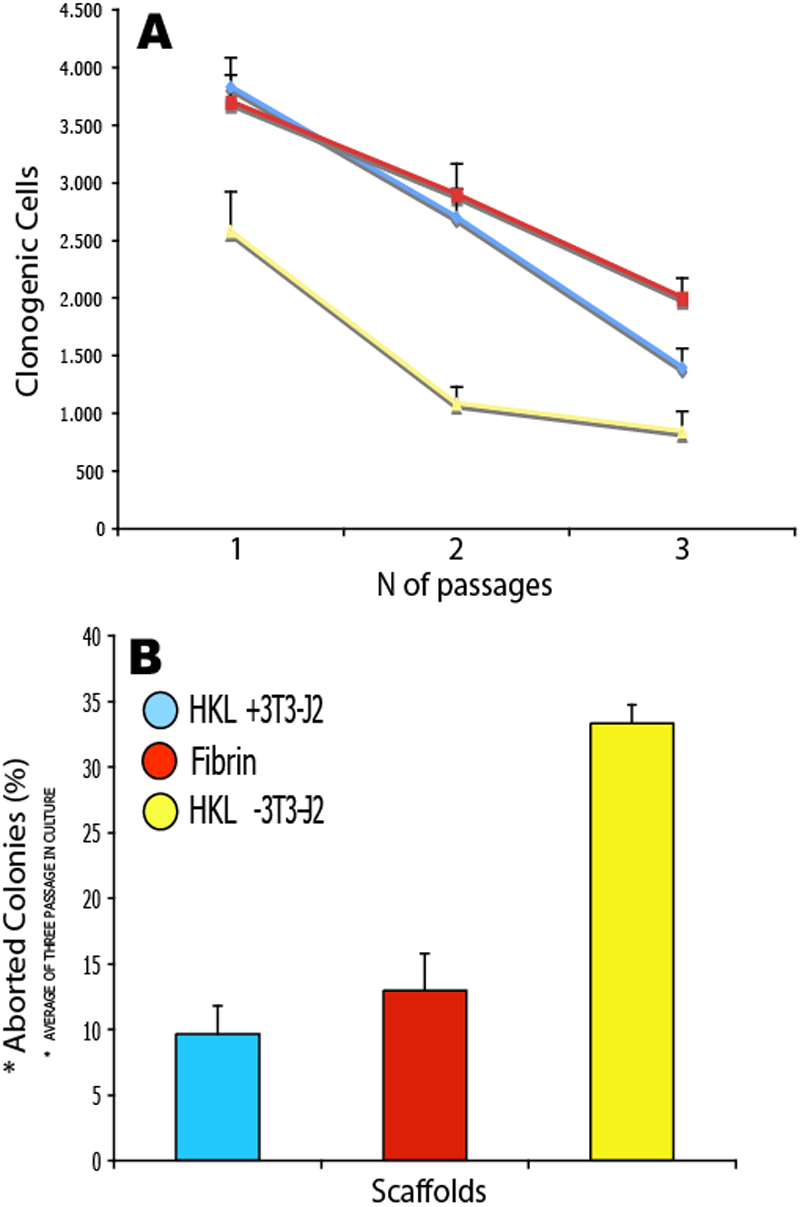
 Figure 5 of Barbaro, Mol Vis 2009; 15:2084-2093.