Figure 2. Comparison between HKL and fibrin-glue matrix as scaffolds for cultured human keratinocytes. Human corneal epithelial stem
cells were cultured onto HKL (A, C) and fibrin glue (B, D) in submerged conditions for 7 days (A, B) and at the air–liquid interface for 14 further days (C, D). Increased stratification (more than 15 cell layers) and overexpression of p63 isoforms in suprabasal cells were only observed
when the fibrin glue was analyzed (D). The growth onto HKL showed a phenotype that was more similar to that of normal human corneal epithelia (A, C). Note that the basal epithelial plane became undulated, yielding an appearance that resembles the palisade of Vogt (bottom
inset, C). Scale bar=50 μm.
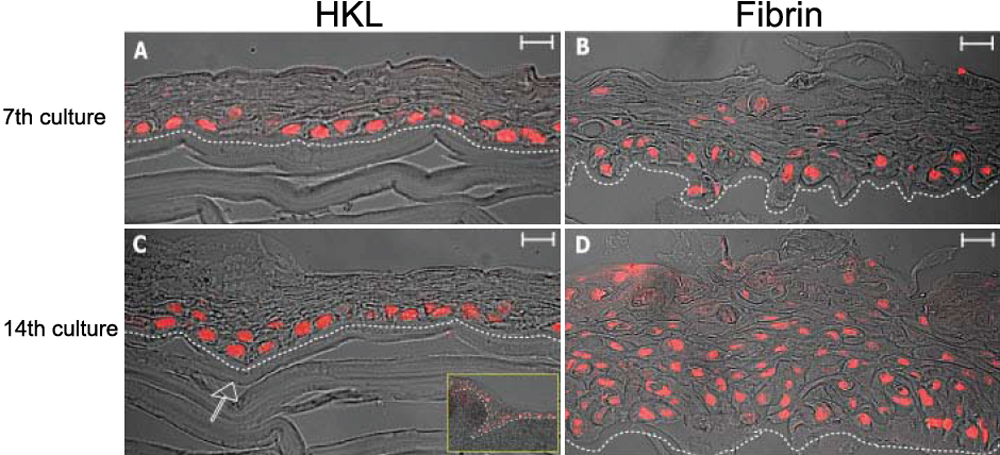
 Figure 2 of
Barbaro, Mol Vis 2009; 15:2084-2093.
Figure 2 of
Barbaro, Mol Vis 2009; 15:2084-2093.