Figure 4. Coronal cerebral T2-weighted
magnetic resonance images. A: Patient I-1 from Family 3. Dashed
arrow shows severe hypogenesis of the corpus callosum with small amount
of remnant tissue localized at the virtual connection between the genu
and the body of the corpus callosum; arrow head shows the atrophic
optic chiasm. B: Normal MRI images. Dashed arrow shows normal
corpus callosum and arrow head shows normal optic chiasm. C:
Patient I-1 from Family 3. Dashed arrow shows lateral callosal bundles
of Probst, which are hemispheric connection fibers that did not cross
the midline and that are seen in callosal dysgenesis. Superomedial
margins of the lateral ventricles are indented by the Probst bundles.
Arrow head shows remnants of the corpus callosum. Lower arrow shows
normal posterior commissure. D: Normal MRI image with dashed
arrow pointing normal corpus callosum.
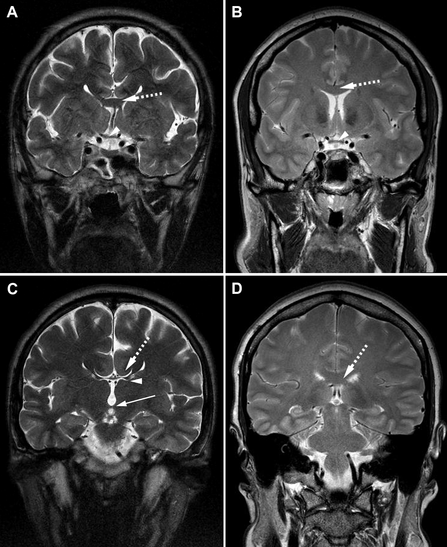
 Figure 4 of Abouzeid, Mol Vis 2009; 15:2074-2083.