Figure 3. Western blot analysis of trabecular meshwork proteins. TM tissue extract (10 µg) was fractionated on a 4–20% gradient gel
(Invitrogen, Carlsbad, CA). A: Western blot analysis after transfer onto a polyvinylidene fluoride membrane. The blot was probed with 17–20 µg of chicken
polyclonal antibody hSRPX#3 (generated against a synthetic peptide; see methods). B: Western blot analysis in normal controls with a protein load of 80 µg and probed with 17–20 µg of chicken polyclonal antibody
hSRPX#3. C: Identical gel as in A stained with Commassie blue R-250.
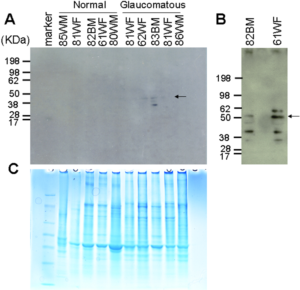
 Figure 3 of
Iragavarapu, Mol Vis 2009; 15:2061-2067.
Figure 3 of
Iragavarapu, Mol Vis 2009; 15:2061-2067.