Figure 6. Cu2+-induced
structural changes of αA- and G98R αA-crystallin. Far-UV (A) and
near-UV (B) CD spectra of 1 mg/ml of αA-crystallin (curve 1) and
G98R αA-crystallin (curve 2) and of 150 μM Cu2+-treated
samples of αA-crystallin (curve 3) and G98R αA-crystallin (curve 4) in
buffer B are shown. C: Changes in the mean hydrodynamic radii (Rh)
of 0.5 mg/ml αA-crystallin and G98R αA-crystallin in the absence (open
bars) and in the presence of 75 μM of Cu2+ (filled bars)
were determined by dynamic light scattering studies. The error bars
represent the statistical variations of the mean hydrodynamic radii of
αA-crystallin or mutant αA-crystallin between 10 experimental data.
G98R αA-crystallin is more susceptible to the Cu2+-induced
structural changes compared to αA-crystallin. [θ]MRM, mean
residue mass ellipticity.
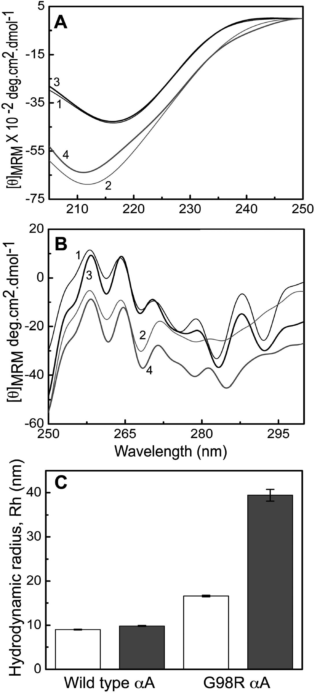
 Figure 6 of Singh, Mol Vis 2009; 15:2050-2060.