Figure 3. Redox-silencing of Cu2+
by αA-crystallin and G98R αA-crystallin. A: Cu2+-ascorbate-mediated
OH˙ generation in the absence (curve 1) and in the presence of
0.05, 0.1, 0.15, 0.25, and 0.4 μM αA-crystallin (curves 2–6). Curve 7
shows the trace of blank sample (in absence of protein and Cu2+).
B: A decrease in coumarin fluorescence intensity (reflecting the
inhibition of OH˙ generation) is shown after 41.6 min as a
function of concentration of αA- (○) and G98RαA-crystallin (●). Inset
shows the percent decrease in fluorescence in the presence of 3 μg/ml
α-crystallins, α-synuclein (Syn), and thyroglobulin (Thy). The results
indicate that G98R mutation in αA-crystallin does not affect its
redox-silencing property. Error bars for four experiments are also
shown.
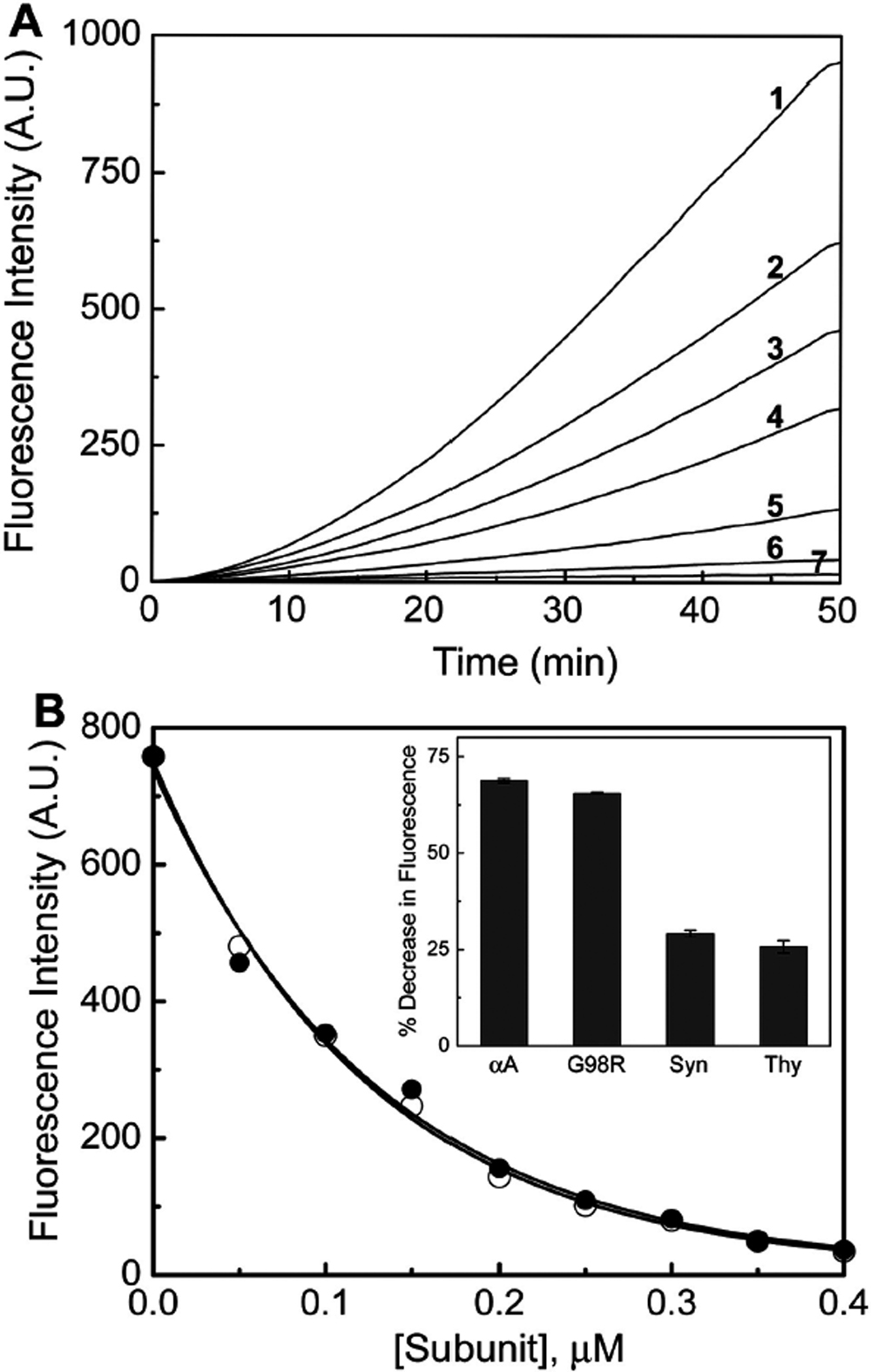
 Figure 3 of Singh, Mol Vis 2009; 15:2050-2060.