Figure 6. Model for visual cycle function.
The different distributions reported in this study led to a model for
visual cycle function. all-trans-Retinol, released from rod
photoreceptor cells during illumination, is taken up via the apical
surface of RPE cells. In turn, 11-cis-retinal is released from
the apical surface for regeneration of rhodopsin. Visual cycle enzymes
convert all-trans-retinol to 11-cis-retinal within the
cell body. The presence of CRALBP and CRBP in both the cell body and
apical processes allows these retinoid-binding proteins to facilitate
diffusion of 11-cis-retinal and all-trans-retinol from
sites of release and uptake, respectively, in the apical processes to
and from enzymatic processing in the cell body. Within the apical
processes, NHERF1, ezrin, and actin interact with CRALBP. Release of 11-cis-retinal
occurs by an unknown mechanism but may be related to complex formation
of interaction of CRALBP, ezrin, and EBP50/NHERF1 in the apical
processes. Release and uptake of retinoids occurs along the whole
apical membrane. It is depicted only from the apical processes for
clarity. CRALBP and CRBP facilitate diffusion by providing binding
sites for retinoids, whose diffusions are driven in either scleral or
vitreal directions by concentration gradients generated by
esterification of retinol by LRAT and perhaps affinity of opsin for 11-cis-retinal,
respectively.
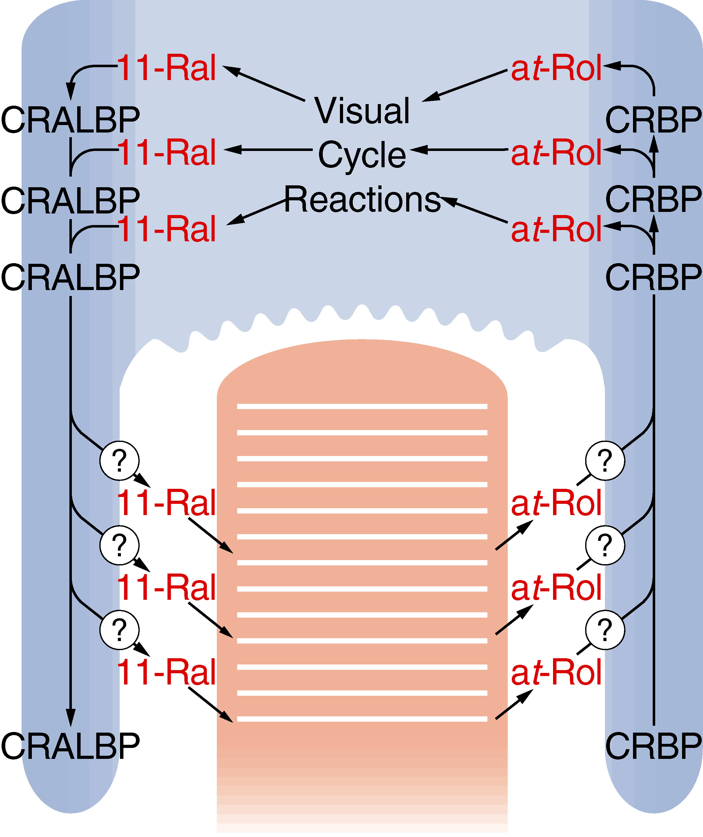
 Figure 6 of Huang, Mol Vis 2009; 15:223-234.