Figure 4. Light and dark experiments. The results illustrate the observed immunocytochemical localization of LRAT (A and B), RHD5 (C and D), and RPE65 (E and F) in retinas from dark-adapted (DA) mice (A, C, and E) and in retinas from mice exposed to room illumination (light-adapted, LA; B, D, F). Dark adapted (DA) mice were held in the dark for 16 h before sacrifice. Light adapted (LA) mice were held in the dark for
16 h, exposed to room illumination for 60 min, and sacrificed.
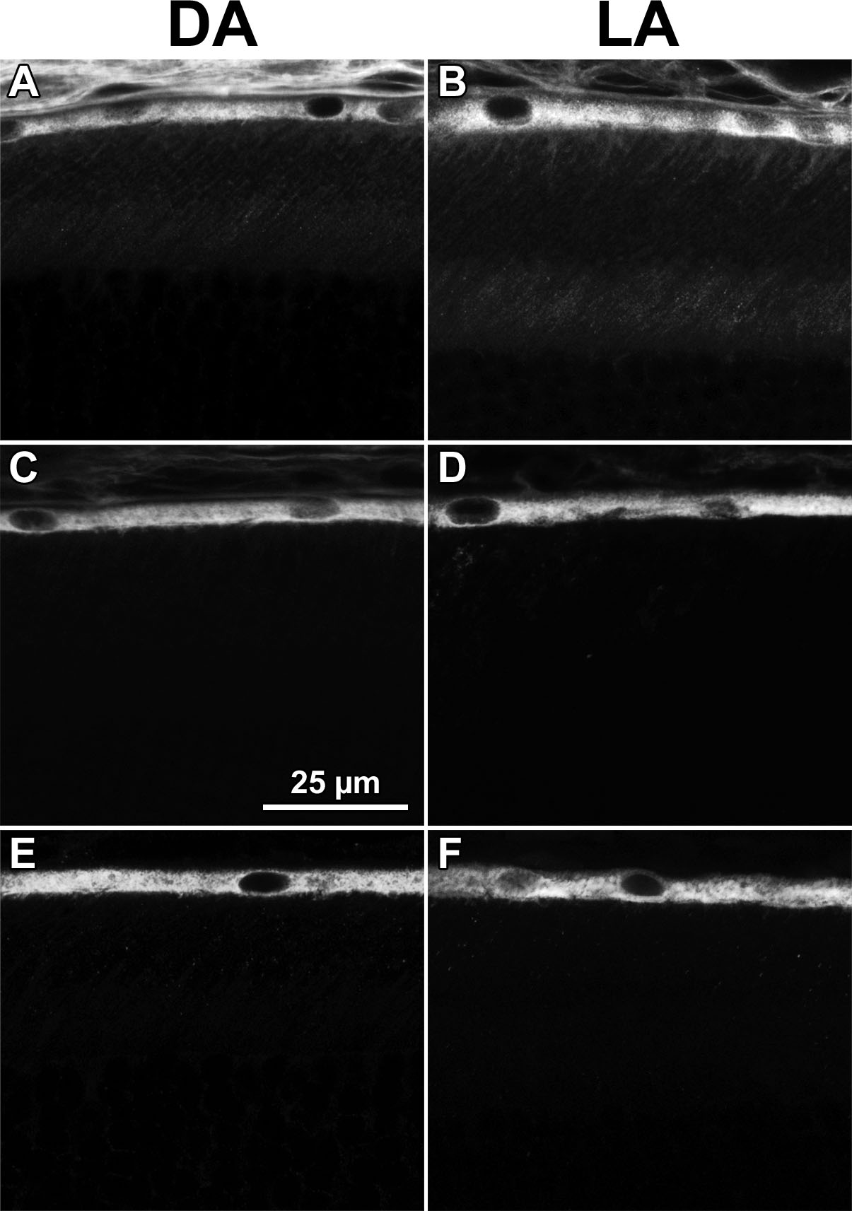
 Figure 4 of
Huang, Mol Vis 2009; 15:223-234.
Figure 4 of
Huang, Mol Vis 2009; 15:223-234.