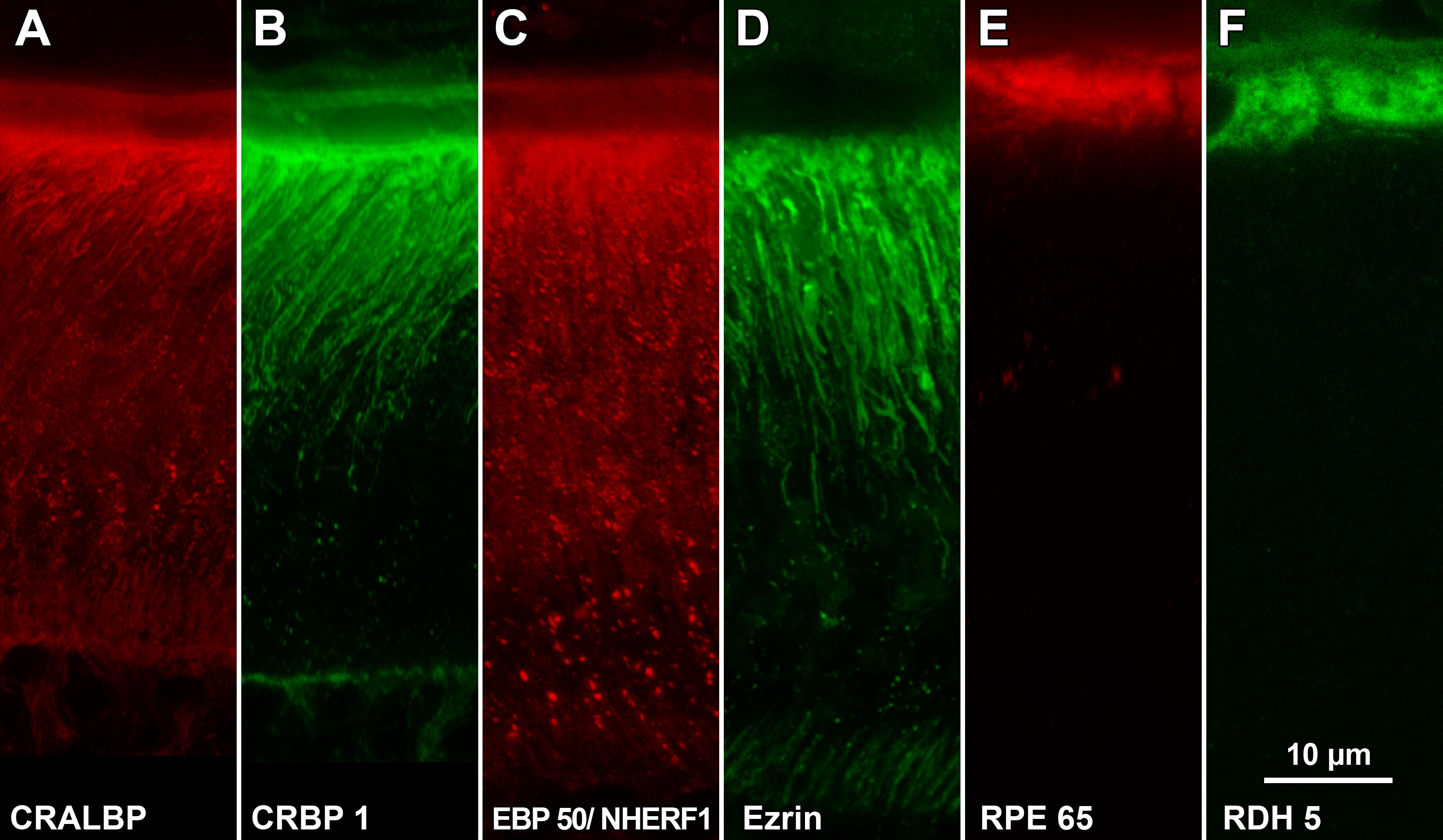
Figure 3. Immunostaining of albino rat
retina. Sections were labeled with fluorescent probes and analyzed by
laser scanning confocal microscopy. The region of outer retina in these
images is similar to that of mouse retina shown in
Figure 1A:
CRALBP is found within the whole RPE cells including the soma and
apical processes (pAb UW55).
B: The distribution of CRBP1 is
similar to that of CRALBP (mAb F3).
C: EBP50/NHERF1 is found
within the soma but primarily within the apical processes (pAb B62).
D:
Ezrin is found primarily within the apical processes (pAb B64).
E:
RPE65 is found within the soma and not within the apical processes (pAb
PETLET).
F: RDH5 is found within the soma and not within the
apical processes (mAb D9). Images shown are Z-projections created from
LSCM image stacks (Z
t=3 μm).
 Figure 3 of Huang, Mol Vis 2009; 15:223-234.