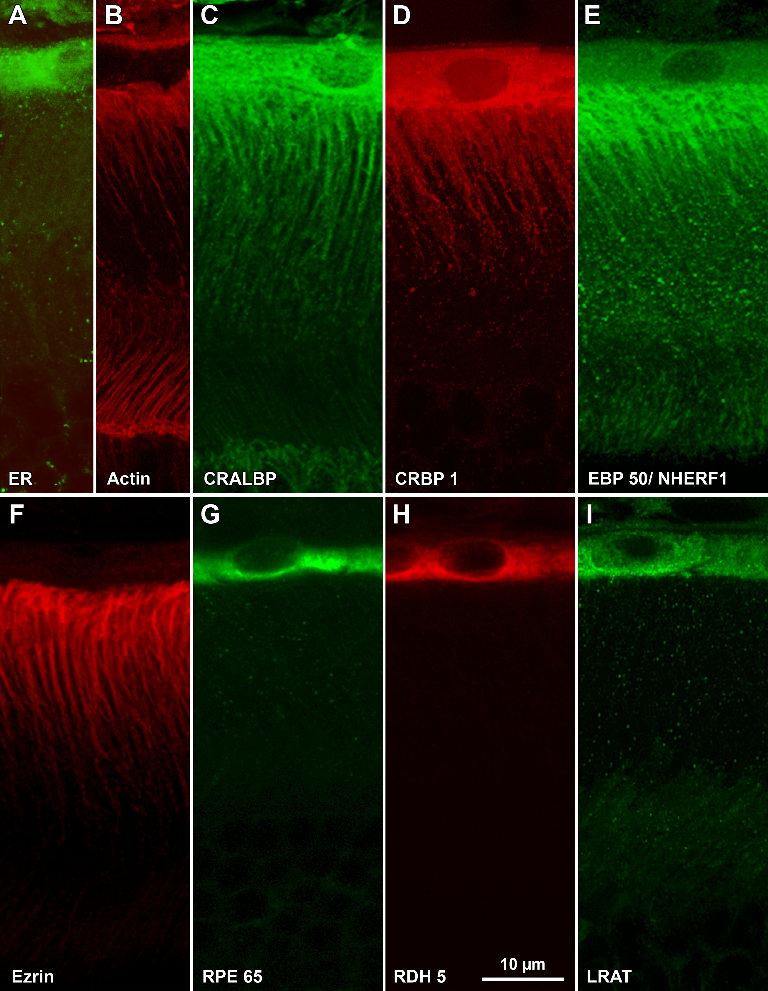
Figure 2. Immunostaining of albino mouse
outer retina. Sections were labeled with fluorescent probes and
analyzed by laser scanning confocal microscopy. The region of outer
retina in these images is shown in
Figure 1A: Calreticulin, a
marker for the endoplasmic reticulum, is found within the RPE cell body
but not its apical processes.
B: Actin, stained with Alexa
Fluor® 488-phalloidin, is found near the basal side of RPE, within RPE
and Müller cell apical processes, and at the external limiting
membrane.
C: CRALBP is found within the entire RPE cell,
including the apical processes. Nuclei appear to be labeled in this
image; however, this was not a consistent feature of the images and is
likely a Z-projection artifact. Distal tips of Muller cells, including
their apical processes, are also labeled (pAb UW55).
D: CRBP1
distribution in RPE cells is similar to that of CRALBP (mAb F3). E.
EBP50/NHERF1 is found within RPE and Müller cell apical processes and,
to a lesser extent, within the RPE cell body. The punctate nature of
the signal has been a constant feature of our images (pAb B64).
F:
Ezrin is confined to RPE apical processes (mAb B62).
G: RPE65
is found within the cell bodies of RPE cells (pAb PETLET). Apical
processes are not labeled.
H: RDH5 distribution is similar to
that of RPE65 (mAb D9).
I: LRAT distribution is similar to
those of RPE65 and RDH5 (mAb B1). Images shown are Z-projections
created from LSCM image stacks (Z
t=3 μm).
 Figure 2 of Huang, Mol Vis 2009; 15:223-234.