Figure 3. NGF modulates TLR4/TLR9 expression in VKC conjunctival epithelial cells (VKC-ECs). A: Relative TLR4/TLR9 transcript expression in few passages VKC-ECs (P1-2). Short-term 1–250 ng/ml NGF exposure induced a steady TLR4 and a slight TLR9 increasing response (p<0.01, REST-ANOVA Tukey–Kramer-coupled analysis). As depicted, the maximum effect was observed at 100
ng/ml NGF, while its specific pretreatment with neutralizing anti-NGF antibody resulted in a downregulation of TLR9 transcripts. Single Ct values were normalized to referring genes, and folds were calculated with respect to their untreated
sister cells. B: Confocal analysis specific for TLR4 (middle) and TLR9 (right) in 100 ng/ml NGF-treated VKC-ECs. Control-isotype signal (left)
was used in channel series acquisitions, and identical acquisition settings were done for all images (60× oil immersion).
C: FACS showed a significant upregulation of both TLR4/TLR9 proteins (5,000 events, right). Note the absence of TLR9 signal
in untreated VKC-ECs (left). TLR2 signal was used as internal control since no difference was observed in molecular analysis.
Isotype-matched control antibody staining was in 101 log decade (IF=7.65; solid area). The graphs are representative of three independent experiments for each sample, which gave
identical results.
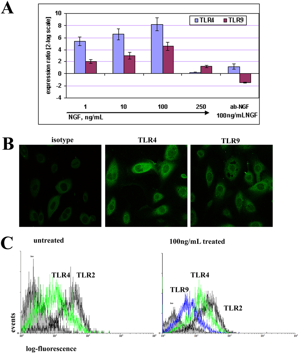
 Figure 3 of
Micera, Mol Vis 2009; 15:2037-2044.
Figure 3 of
Micera, Mol Vis 2009; 15:2037-2044.