Figure 1. Characterization of primary conjunctival epithelial cells outgrew from VKC explants (VKC-ECs). A: Phase contrast microscopy (Normasky) showing VKC-ECs migrating from explants after 1 week (left, 10× optic field). The picture
is representative of all VKC biopsies. A representative forward/side scatter plot of gated primary VKC-ECs (middle) and a
FC-CK19 histogram demonstrating the purity of outgrew ECs (right; solid area: background staining (negative control); bold
line: 97% CK19+ ECs). B: Confocal analysis specific for TLR4 and TLR9 (merge) in VKC-ECs. Control-isotype signal (data not shown) was used in channel
series acquisitions, and identical acquisition settings were carried out for all images (60× oil immersion). C: TLR4/TLR9 transcript expression in untouched primary cultures of VKC-ECs (P0; p<0.01, REST-ANOVA Turkey–Kramer-coupled analysis).
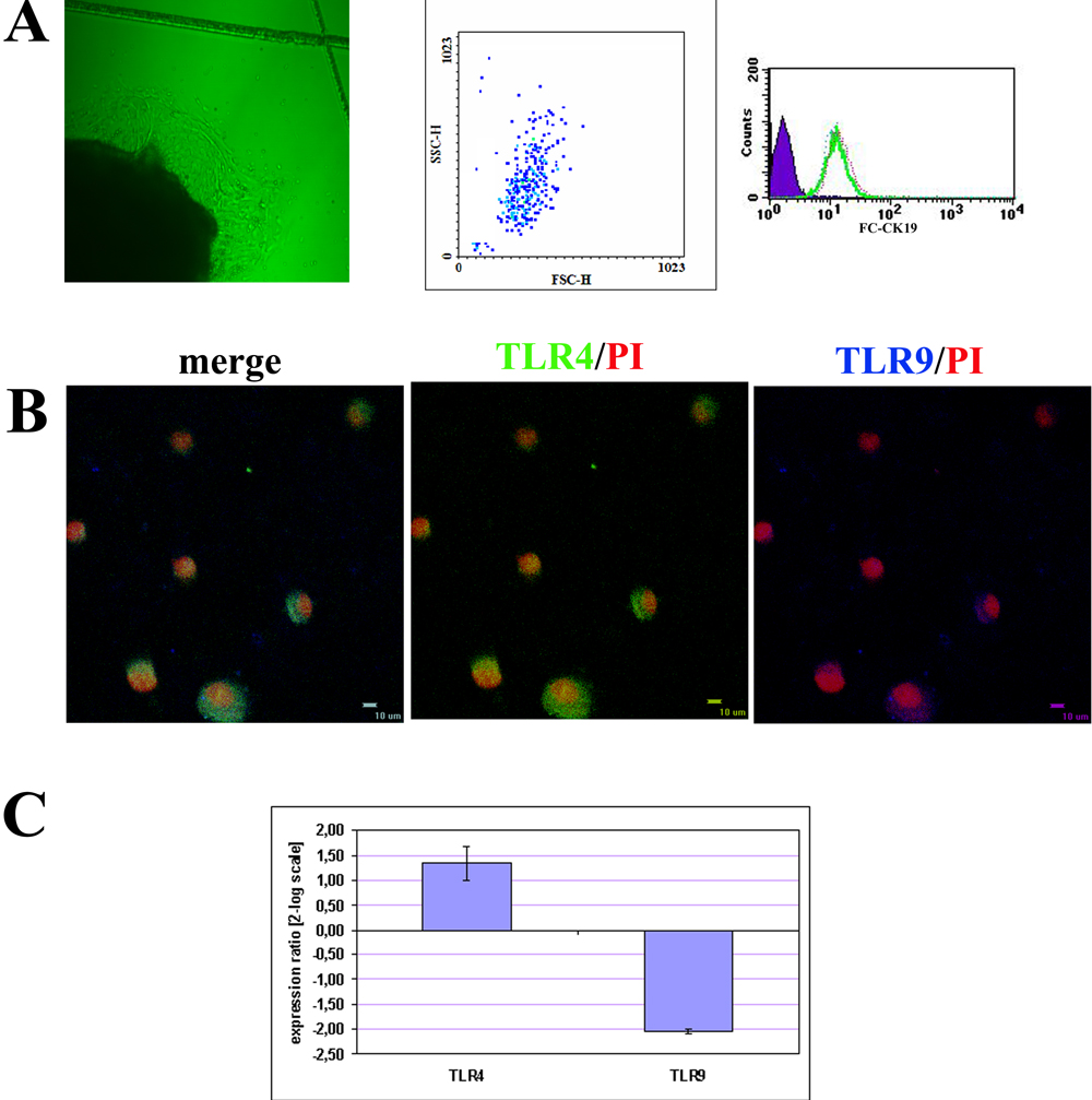
 Figure 1 of
Micera, Mol Vis 2009; 15:2037-2044.
Figure 1 of
Micera, Mol Vis 2009; 15:2037-2044.