Figure 3. Representative photographs and H-E stained sections of the porcine corneas decellularized by various methods. The left column
shows photograph of native cornea (A), cornea treated with Triton® X-100 (C), cornea treated with SDS (E) and cornea decellularized by UHP (G). The right column shows H&E stained section of native cornea (B), cornea treated with Triton® X-100 (D), cornea treated with SDS (F) and cornea decellularized by UHP (H). Epithelial cells and keratocytes are seen in the corneas treated with Triton® X-100 or SDS but not in the cornea treated
with UHP. Scale bar, 50 µm.
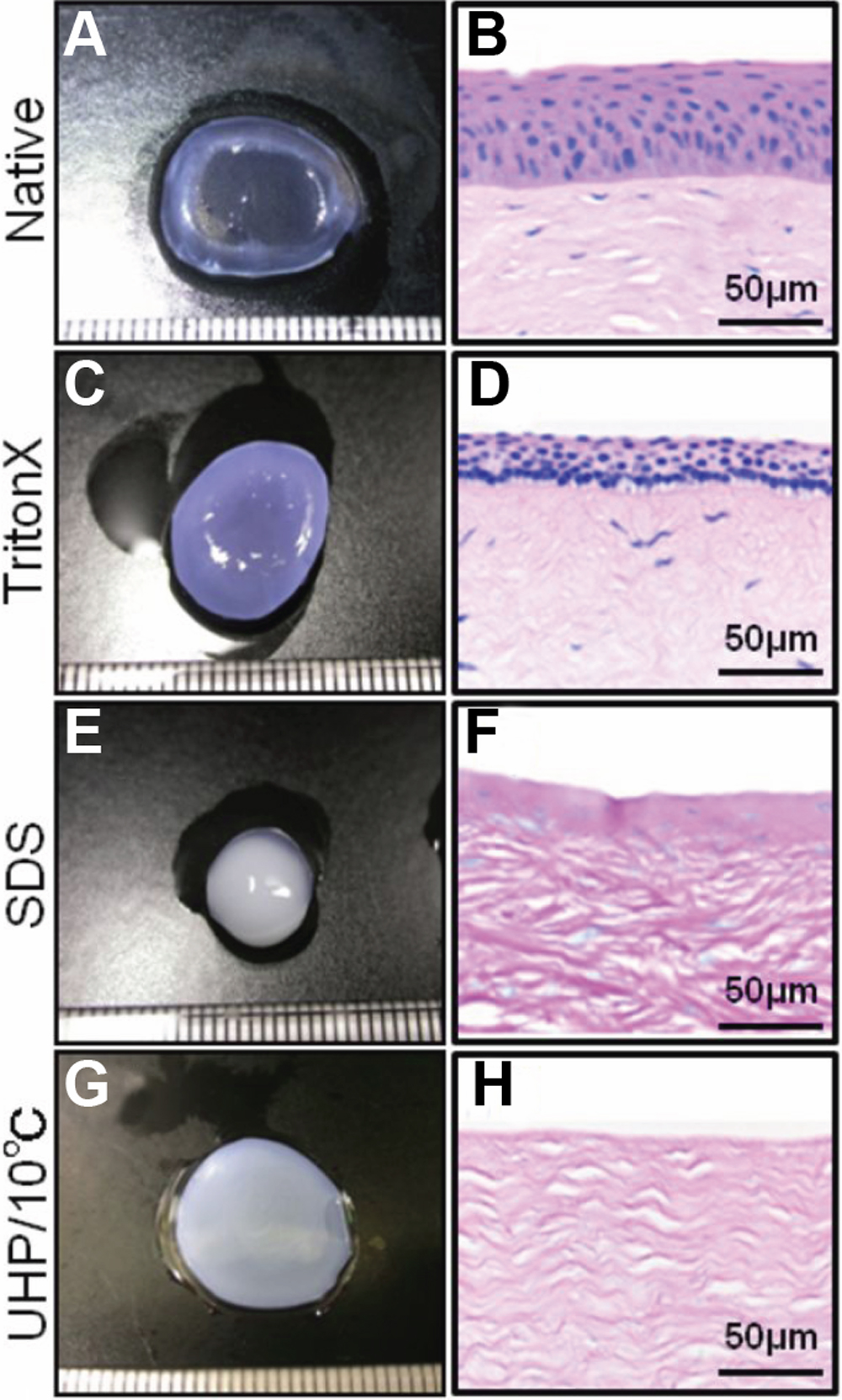
 Figure 3 of
Sasaki, Mol Vis 2009; 15:2022-2028.
Figure 3 of
Sasaki, Mol Vis 2009; 15:2022-2028.