Figure 2. Demonstration of VEGFR1 protein in corneal fibroblasts by immunofluorescence. The presence of VEGFR1 was investigated using
antibodies specific to VEGFR1 and VEGFR2. A: Significant levels of VEGFR1-specific fluorescence in type 1 donor fibroblasts. VEGFR1 immunoreactivity was undetectable
in type 2 donors cells (C). B (type -1) and D (type-2) represent cells treated with anti-VEGFR2 antibodies, providing negative controls.
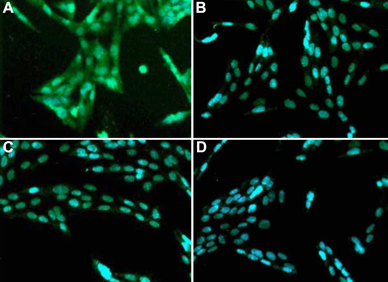
 Figure 2 of
Berthaut, Mol Vis 2009; 15:1997-2007.
Figure 2 of
Berthaut, Mol Vis 2009; 15:1997-2007.