Figure 1. Network formation by corneal fibroblasts on Matrigel. Type-1(A) and type-2 (C) corneal fibroblasts were seeded on matrigel and photographed before incubation, representing time 0. The same plates were
photographed after 2 h of incubation. B (type-1 corneal fibroblast) shows cell elongation and beginning of network formation whereas D (type-2 fibroblast) shows no network formation, and resembles the unincubated controls at time 0.
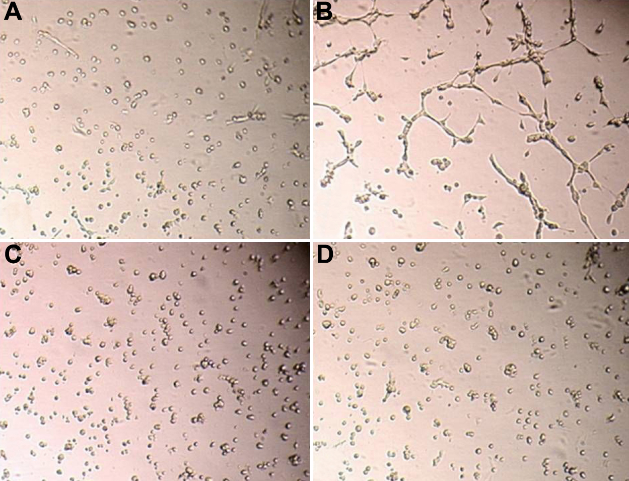
 Figure 1 of
Berthaut, Mol Vis 2009; 15:1997-2007.
Figure 1 of
Berthaut, Mol Vis 2009; 15:1997-2007.