Figure 6. Inhibition of corneal neovascularization by anti-VEGF antibody. Comparison of corneal neovascularization in control (A and C) and anti-VEGF-treated mice (B and D) at 7 days p.i. A: C. albicans keratitis results in multiple blood vessels arising from the limbal arcade and extending toward the central cornea. B: Treatment with VEGF-blocking antibody results in fewer and shorter corneal blood vessels that remain limited to the peripheral
cornea. C: Image analysis of corneal blood vessels in a PBS-treated mouse. D: Image analysis of corneal blood vessels in anti-VEGF-treated mouse, ignoring underlying radial iris vessels that had a slightly
larger caliber but lacked visible iris neovascularization.
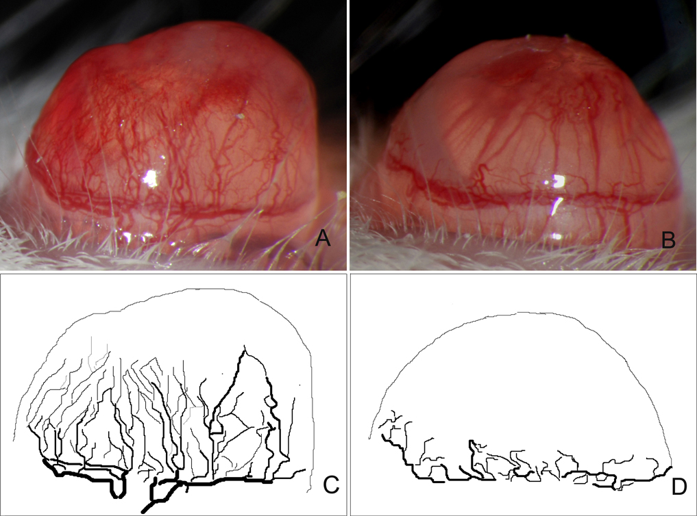
 Figure 6 of
Yuan, Mol Vis 2009; 15:1988-1996.
Figure 6 of
Yuan, Mol Vis 2009; 15:1988-1996.