Figure 3. Molecular expression patterns in situ in corneas with C. albicans infection. VEGF-A expression was compared between corneas with C. albicans keratitis (Infected) and mock-inoculated controls (Mock). Negative controls lacked primary antibody (Control”. Corneal sections
were processed with anti-VEGF-A monoclonal antibodies (VEGF-A). Immunofluorescence showed VEGF-A in the healed epithelium
of scarified corneas and within the epithelium and stroma one day after the onset of experimental C. albicans keratitis. Original magnification, 10×. Scale bar, 20 μm.
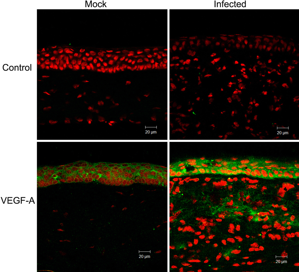
 Figure 3 of
Yuan, Mol Vis 2009; 15:1988-1996.
Figure 3 of
Yuan, Mol Vis 2009; 15:1988-1996.