Figure 1. Identification of HCM cells. The
third passaged HCM cells were washed with PBS, fixed with formaldehyde,
blocked in BSA/PBS and stained with monoclonal anti-smooth muscle
α-actin and rhodamine-conjugated goat anti-rabbit IgG;HCM cells were
also stained with monoclonal rabbit anti-Desmin and goat anti-rabbit
IgG. HCM cells labeled with antibody to smooth muscle actin (A;
red) and the negative control (B). Nuclei staining with DAPI is
shown in blue. HCM cells labeled with antibody to desmin (Buffy; C)
and the negative control (D).
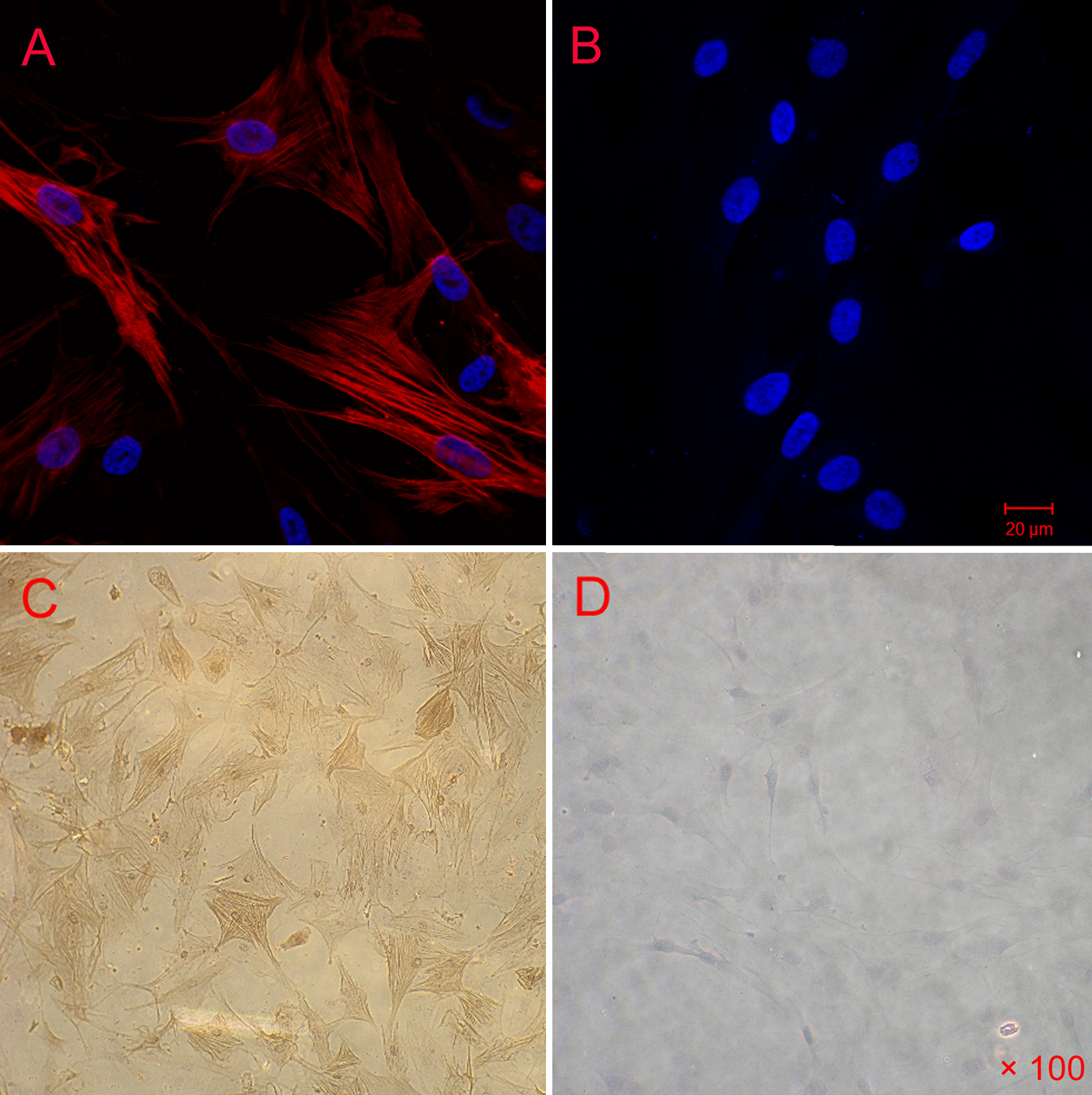
 Figure 1 of Lan, Mol Vis 2009; 15:1977-1987.