Figure 3. Double immunofluorescence showing colocalization of galectin-3 with either glutamine synthetase, or GFAP in six-month-old
pig retinas. Galectin (GC)-3 (A; red fluorescence, arrows) is colocalized in most glutamine synthetase (GS)-positive cells (B; green fluorescence, arrows). The arrows in each figure indicate Müller cell bodies. The colocalization of galectin-3 and
GS is shown in C (merged image). Galectin-3 (D; red fluorescence, arrow) was also observed in GFAP-positive cells (E; green fluorescence, arrow) in the retina. Colocalization is shown in F (merged image). The arrow in each figure (D-F) indicates GFAP-positive processes across all retinal layers. C and F are merged images (yellow fluorescence, arrow) of a six-month-old pig retina. Scale bars represent 50 μm.
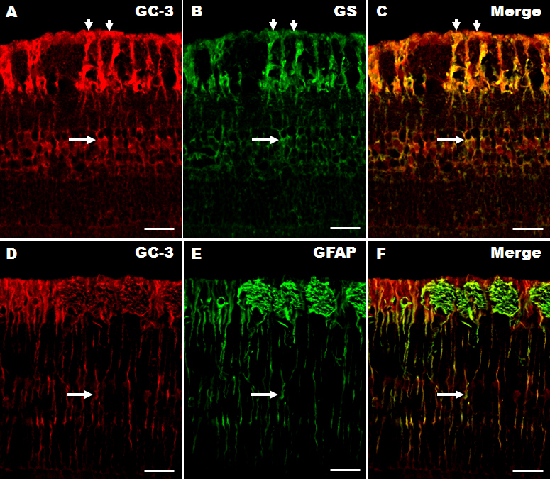
 Figure 3 of
Kim, Mol Vis 2009; 15:1971-1976.
Figure 3 of
Kim, Mol Vis 2009; 15:1971-1976.