Figure 2. Immunohistochemical localization of galectin-3 in two-day-old and six-month-old pig retinas. A: In the retina of a two-day-old pig, galectin-3 immunostaining is seen in some cells in the GCL (arrowheads) and INL (arrows).
Weak galectin-3 immunoreactivity was detected in the INL and outer limiting membrane. B: In the retina of a six-month-old pig, the pattern of galectin-3 immunoreactivity was similar to that seen in the retina
of the two-day-old pig shown in (A). Galectin-3 was immunostained in glial cell processes from the nerve fiber layer to the outer limiting membrane. Particularly,
the immunoreactivity in the GCL (arrowheads) and INL (arrows) was enhanced, compared with that in the two-day-old pig. C: This is a negative control (two-day-old pig retina). No specific reaction product is seen in sections incubated with nonimmune
serum. Abbreviations: ganglion cell layer (GCL); inner plexiform layer (IPL); inner nuclear layer (INL); outer plexiform layer
(OPL); outer nuclear layer (ONL); rod and cone layer (RCL). A–C were counterstained with hematoxylin. Scale bars represent 50 μm.
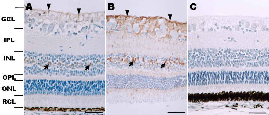
 Figure 2 of
Kim, Mol Vis 2009; 15:1971-1976.
Figure 2 of
Kim, Mol Vis 2009; 15:1971-1976.