Figure 5. Comparison of the effect of denuded HAM by means of an enzyme, Dispase and mechanical scraping of epithelial cells after incubation.
Left panel: Transmission electron micrograph showing the effect of different incubations with Dispase for 30, 60, or 120 min
on HAM. After 30 min incubation with Dispase, basal cells of the remnant epithelium sent out cytoplasmic blebs. After 60 min,
the lamina densa was disrupted with exposure of stromal collagen. After 120 min, further loosening of the collagen structure
occurred. Magnification: 30, 000×, the scale bar indicates 0.5µm. Right panel: Scanning electron micrograph showing the surface
of denuded HAM by Dispase. Longer incubations produced a smoother surface with a more open collagen structure.
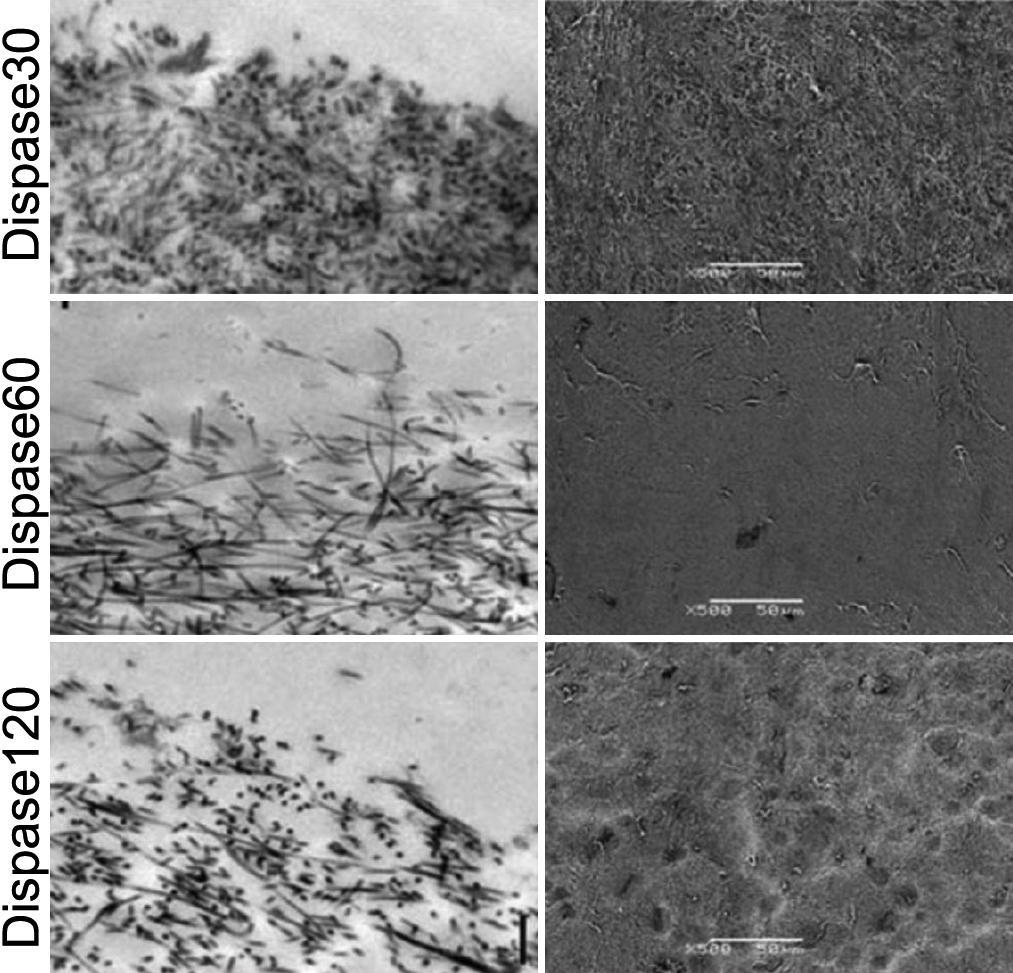
 Figure 5 of
Lim, Mol Vis 2009; 15:1962-1970.
Figure 5 of
Lim, Mol Vis 2009; 15:1962-1970.