Figure 4. Transmission electron micrograph of untreated HAM tissue (normal control). The basement membrane supports the overlaying epithelium
and is composed of a three-layered basal lamina and lamina fibroreticularies. Lamina rara externa (lucida) is an electron-lucent
zone directly bordering the adjacent cell which makes up the upper portion of the basal lamina. Lamina densa (LD) is an electron-dense
zone that appears somewhat amorphous and granular, and constitutes the intermediate part of the basal lamina. Lamina rara
interna comprises the basal portion of the basal lamina. The three layers of basal lamina sit on top of the lamina fibroreticularies,
which is synthesized by cells from underlying connective tissue and contains fibrillar structures namely anchoring fibrils,
elastic fibrils and microfibril bundles. In the image, “E” indicates epithelium, “LD” indicates lamina densa, and “S” indicates
stroma. Magnification: 30, 000×, the scale bar indicates 0.5µm.
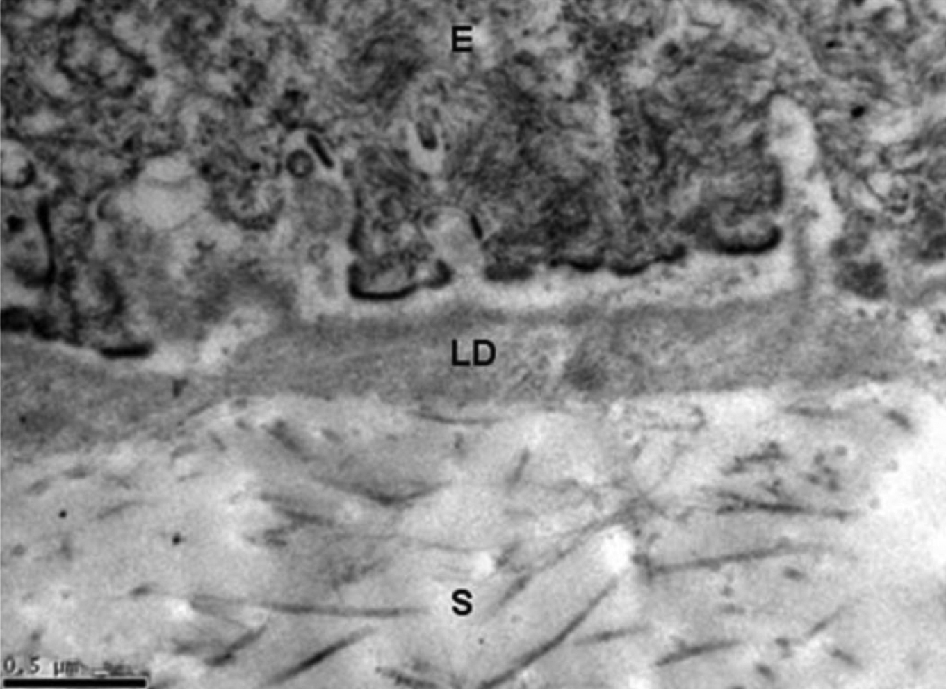
 Figure 4 of
Lim, Mol Vis 2009; 15:1962-1970.
Figure 4 of
Lim, Mol Vis 2009; 15:1962-1970.