Figure 2. Histology of chemically burned corneas stained with H&E. H&E staining shows much more new vessels and mononuclear cells presented
in control corneal stroma compared with PSP treated cornea. Three sections from different mice are shown in representative
micrographs. Bar, 50 µm.
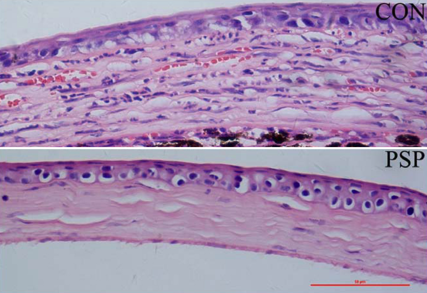
 Figure 2 of
Yang, Mol Vis 2009; 15:1951-1961.
Figure 2 of
Yang, Mol Vis 2009; 15:1951-1961.