Figure 1. Macroscopic observation of corneal neovascularization in PSP-treated and control mice. PSP (100 μg/ml) or solvent control
was used topically 4 times everyday on alkali-injured mice cornea for 7 days, images were taken with slit lamp. It can been
seen clearly that in control group without PSP treated, the new vessel is predominant in conea, however which is very slight
in cornea treated with PSP.
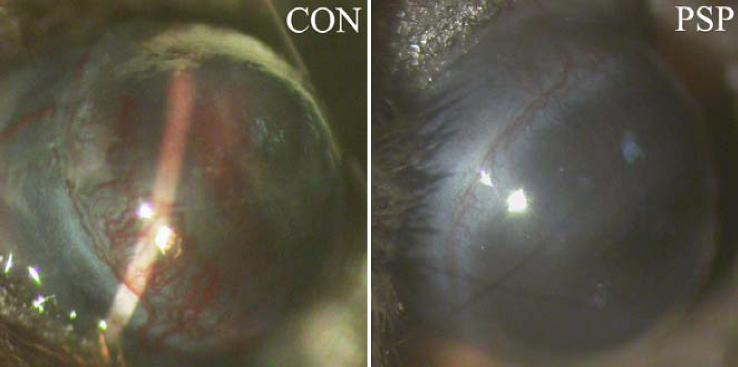
 Figure 1 of
Yang, Mol Vis 2009; 15:1951-1961.
Figure 1 of
Yang, Mol Vis 2009; 15:1951-1961.