Figure 1. Sequence of the region of the mutation. MYOC sequence electropherogram is shown of a wild-type unaffected subject (top panel) and of a heterozygous patient carrying
the myocilin mutation, Y371D (lower panel). Nucleotides and predicted amino acid changes are indicated under the electropherogram.
The vertical line points to the Y371D mutation.
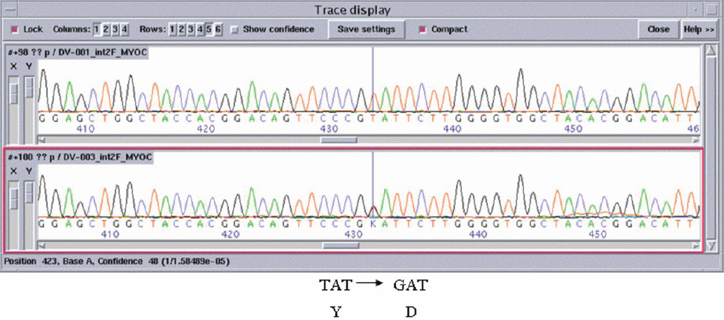
 Figure 1 of
Avisar, Mol Vis 2009; 15:1945-1950.
Figure 1 of
Avisar, Mol Vis 2009; 15:1945-1950.