Figure 6. Pig retinal cryosections labeled immunohistochemicaly for photoreceptor protein markers. A: Cells in the prospective ONL are positive for recoverin (arrows) in GW9 retina. ONL and photoreceptor segments are recoverin-labeled
in 1W (B) and 6M (C) retina. D: Some differentiating rod photoreceptors are positive for rhodopsin in GW9 retina (arrows). E: Strong rhodopsin-labeling is present in the outer segments (thick arrows) in 1W retina. Inner segments (arrowheads) and
cell bodies (thin arrows) are less intensely stained. F: Rhodopsin-positive inner (arrowheads) and outer (thick arrows) segments are more elongated in 6M compared to 1W retina.
Some rod cells bodies are also labeled (thin arrows) in 6M retina. Cone outer segments are labeled for GNAT2 in 1W (G) and
6M (H) retina (thin arrows). Nuclei are labeled with PI (red).
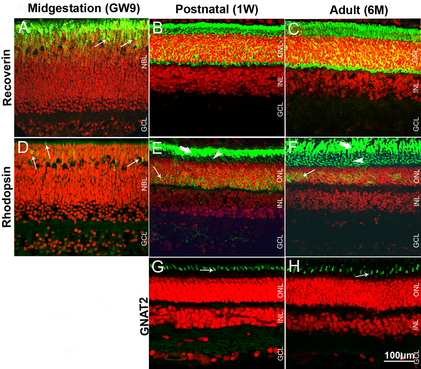
 Figure 6 of
Guduric-Fuchs, Mol Vis 2009; 15:1915-1928.
Figure 6 of
Guduric-Fuchs, Mol Vis 2009; 15:1915-1928.