Figure 1. Pig retinal cryosections labeled immunohistochemicaly for nestin, Pax6 and Ki67. A: Nestin immunoreactivity is evident throughout GW9 retina. B: Nestin positive fibers are in the GCL and IPL (arrows) in 1W retina. C: Cells in the GCL, differentiating amacrine (thick arrows), horizontal (arrowheads) and putative retinal progenitors (thin
arrows) are Pax6-positive in GW9 retina. D: Immunoreactivity for the mitotic marker Ki67 in GW9 retina reveals distribution of progenitor cells similar to that labeled
for Pax6 (thin arrows). E: Cells in the GCL (thick arrows), amacrine (thin arrows) and some horizontal cells (arrowheads) are Pax6-positive in 1W retina.
Nuclei are labeled with PI (red).
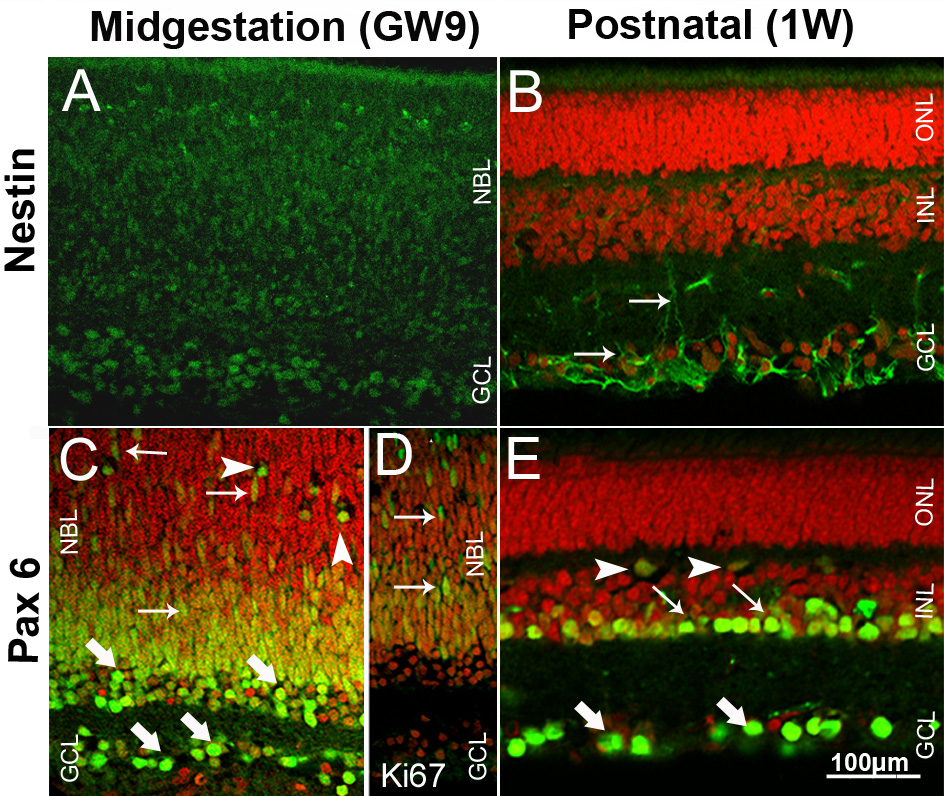
 Figure 1 of
Guduric-Fuchs, Mol Vis 2009; 15:1915-1928.
Figure 1 of
Guduric-Fuchs, Mol Vis 2009; 15:1915-1928.