Figure 6. Immunofluorescence analysis of choroidal vascularity in rat eyes after topical VS application. The choroidal vascularity in
PBS-treated or VS-treated eyes was examined by immunofluorescence analysis using anti-vWF. A: Profile of vWF-positive vessels in choroids (C) and retina (R) of PBS-treated or VS-treated eyes are shown. Arrows indicate
CNV by little circles in low power filed (arrowheads) and big circles in high power field (larger arrows). B: PBS-treated vWF-positive blood vessels in choroids are nearly twice those of VS-treated eyes. Data are summarized as mean±SEM
(n=8). Asterisk (*) indicates p<0.05. The scale bar equals 200 μm.
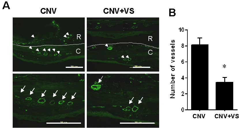
 Figure 6 of
Sheu, Mol Vis 2009; 15:1897-1905.
Figure 6 of
Sheu, Mol Vis 2009; 15:1897-1905.