Figure 5. Histological analysis of CNV lesions in rat eyes after topical VS application. After final FAG analysis on day 42, rat eyes
were dissected and analyzed by hematoxylin and eosin analysis. VS application resulted in much smaller neovascularization.
Arrows indicate the laser-induced CNV lesions. Abbreviations: ganglion cell layer (GL); inner nuclear layer (INL); outer nuclear
layer (ONL); choroid (CH). Magnification is 100×.
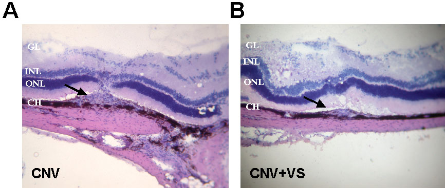
 Figure 5 of
Sheu, Mol Vis 2009; 15:1897-1905.
Figure 5 of
Sheu, Mol Vis 2009; 15:1897-1905.