Figure 4. Effect of topical VS application on CNV lesions in rats by FAG. A: Representative photographs of fundus microscopy (top panels) and FAG (bottom panels) indicate the anti-angiogenic effect
of topical VS application. Top: the laser spots in rat eyes of different group are shown under fundus microscope. Bottom:
FAG analysis of CNV lesion in rat eyes by group are shown on day 28. B: Effect of topical VS application on the area of CNV lesions in rats are evaluated by FAG. Areas of CNV lesions were determined
by FAG examination on days 21, 28, 35, and 42 after laser photocoagulation. Data are summarized as mean±SEM (n=8) and are
representative of three experiments. Asterisk (*) represents p<0.05, and double asterisk (**) represents p<0.01.
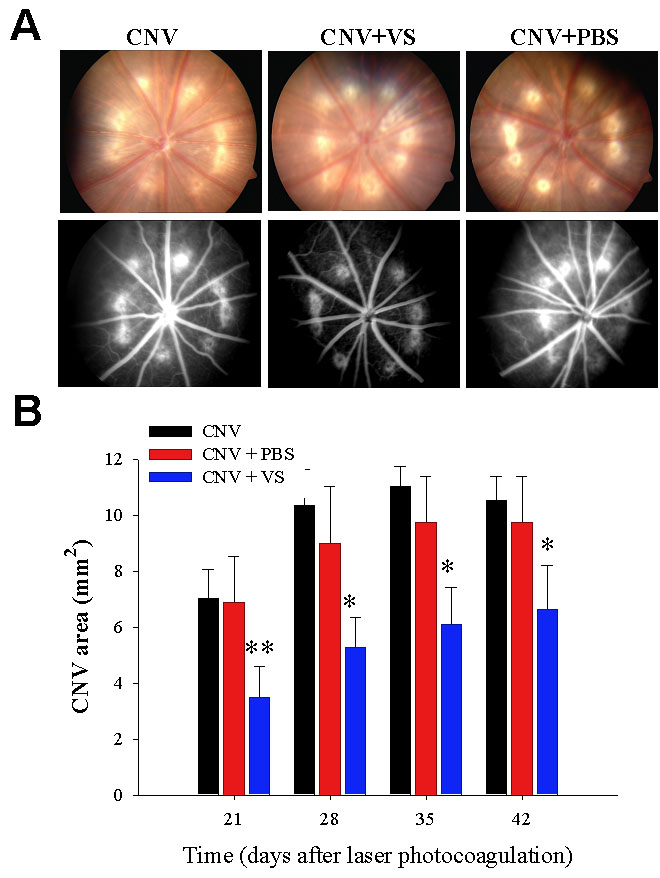
 Figure 4 of
Sheu, Mol Vis 2009; 15:1897-1905.
Figure 4 of
Sheu, Mol Vis 2009; 15:1897-1905.