Figure 1. Anti-angiogenic function of VS in endothelial cells. A: Effect of VS on migration of HUVECs. VS was added in varying doses, and endothelial cells were placed in a Boyden chamber
for 6 h and allowed to migrate toward bFGF. Shown are representative photographs of endothelial migration in control and after
treatment with 1 μg/ml VS (left). Cell migration was quantified by counting cells from three high power fields and expressed
as mean ± SD of triplicates (right). B: Effect of VS on tube formation of HUVECs. Endothelial cells were applied to Matrigel and incubated for 4 h in the presence
of VS of varying doses. Representative profiles of the tubular structures in control and 1 μg/ml VS-treated HUVEC are shown
(left). Tube formation was quantified by counting the number of rings and expressed as mean±SD from quadruplicates (right).
Asterisk (*) represents p<0.05, and double asterisk (**) represents p<0.01.
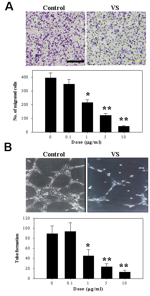
 Figure 1 of
Sheu, Mol Vis 2009; 15:1897-1905.
Figure 1 of
Sheu, Mol Vis 2009; 15:1897-1905.