Figure 6. Laser injury stimulates directed
Müller cell body migration. GFP-positive Müller cells were imaged for
150 min following laser burn injury. A-K shows the same
area every 15 min at day 2 post injury. L is an overlay of the
first (0 min, green) and the last (150 min, red) time point to present
a comparison of the position of the cell bodies directly. The live
imaging data indicated that the cell bodies of selected Müller cells
were translocated 30 μm toward the injury site within those 150 min of
imaging, suggesting that Müller cell bodies are able to migrate with a
speed of approximately 12 μm/h. The scale bar represents 50 μm.
Abbreviations: GCL represents ganglion cell layer.
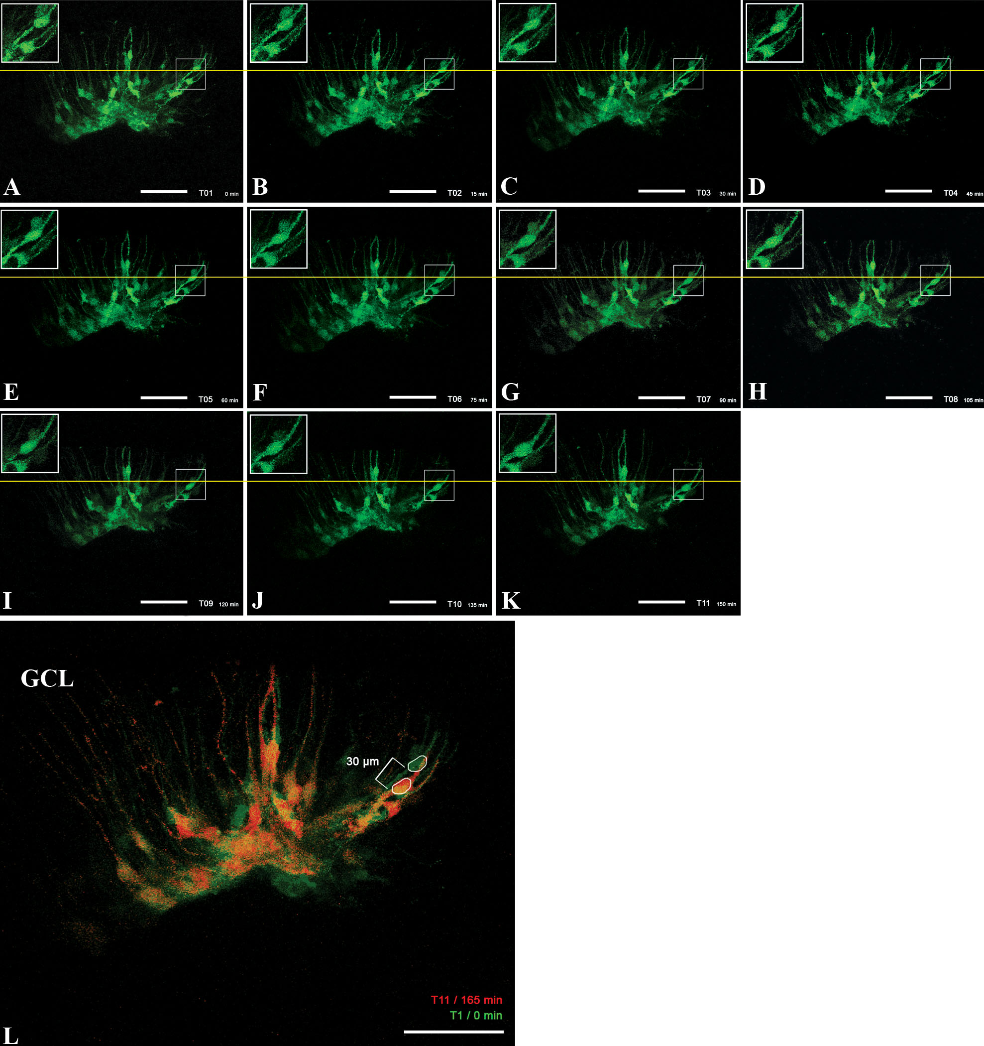
 Figure 6 of Tackenberg, Mol Vis 2009; 15:1886-1896.