Figure 5. Cyclin-D3 positive cell bodies
in the ONL suggested Müller cell migration. Representative examples of
Cyclin D3-positive cell bodies in the outer nuclear layer (ONL) at two
days following laser burn injury, suggesting possible Müller cell
migration. A: The injury site is shown at day 2 post injury,
stained with vimentin (green) and Cyclin D3 (red). B: The
injury site is shown at day 2 post injury, stained with nestin (green)
and Cyclin D3 (red). Arrows indicate Cyclin D3-positive cell bodies in
the ONL localized around the injury site. The scale bar represents 75
μm. Insets are higher power views of the indicated region.
Abbreviations: GCL represents ganglion cell layer.
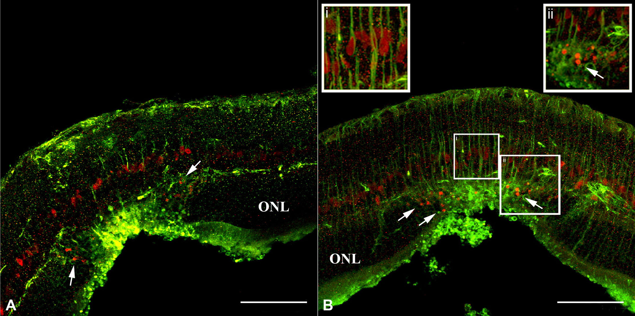
 Figure 5 of Tackenberg, Mol Vis 2009; 15:1886-1896.