Figure 4. Laser injury stimulates Müller
glia proliferation. Antibodies, targeted against GS (A, C;
green), GFAP (B, D; green), Cyclin D1 (A, B;
red), and Cyclin D3 (C, D; red) were applied at two days
postretinal injury. Müller glia within the injury site stain positive
for the cell cycle markers Cyclin D1 and D3, indicating that laser burn
induces a reentry into the cell cycle and subsequent glial cell
proliferation. The scale bar represents 75 μm. Abbreviation: outer
nuclear layer (ONL). Insets are higher power views of the indicated
region. Arrows point to positively labeled nuclei in A, B i,
and ii, C, and D.
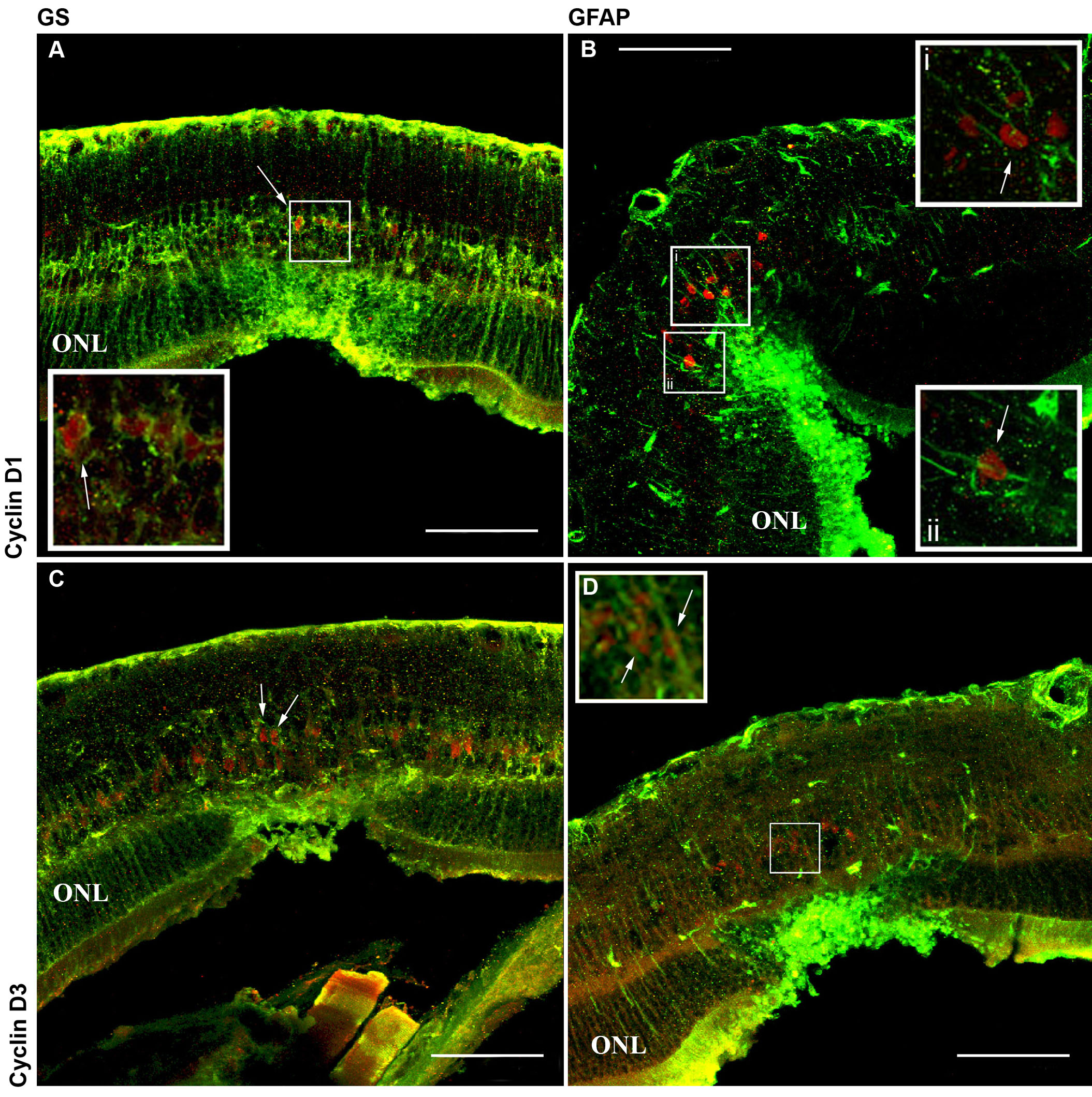
 Figure 4 of Tackenberg, Mol Vis 2009; 15:1886-1896.