Figure 2. Expression of cell cycle
markers. Cell bodies in the inner nuclear layer (INL) reentered the
cell cycle, began to proliferate, and migrated to the ONL following
laser injury. The cell cycle marker Cyclin D1 and the Müller cell
nuclear marker and cell cycle marker Cyclin D3 were used to identify
proliferative cells. Cyclin D1 staining was elevated within 24 h after
laser injury and localized within the injury site only. Cyclin D3
expression was increased at days 2 and 3 postinjury (H,I).
Positive cells normally found in the INL were now located in the outer
nuclear layer, indicated by arrows H and I (ONL; H-J).
The scale bars represent 75 μm.
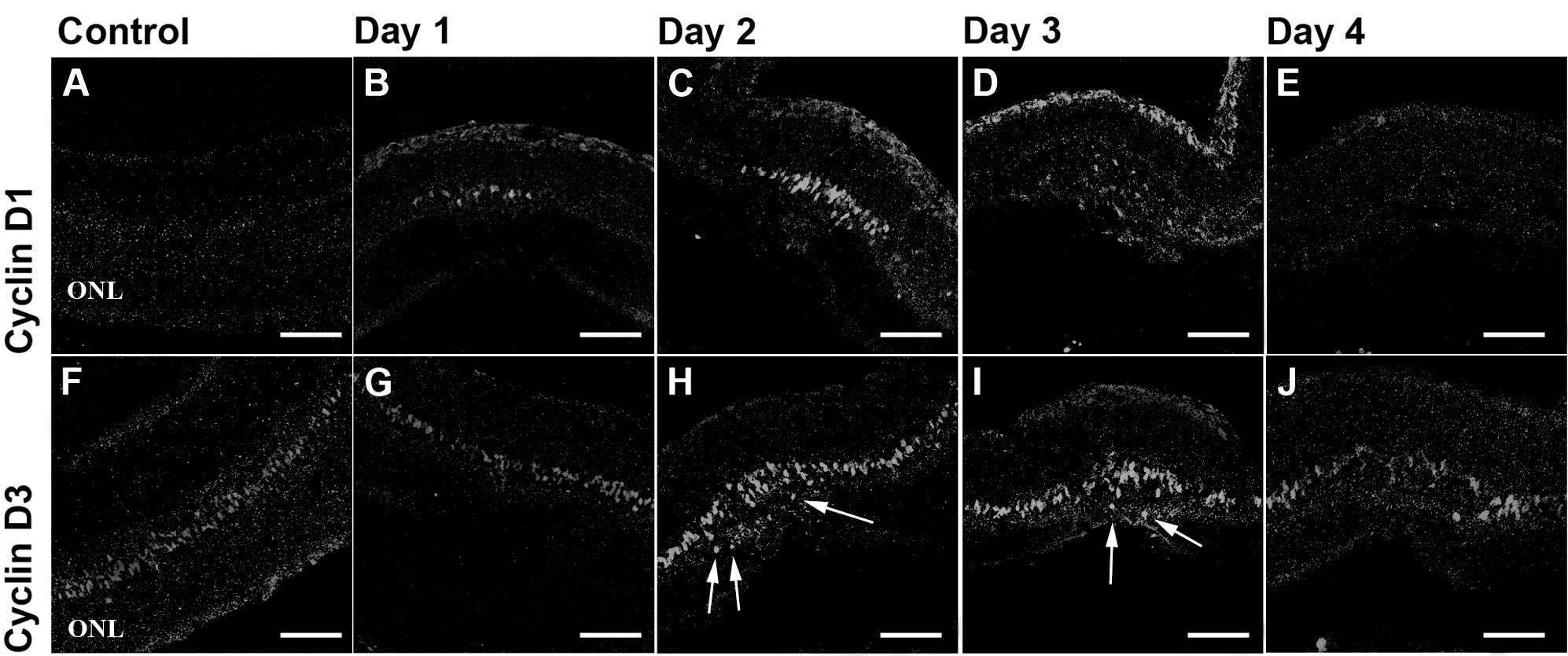
 Figure 2 of Tackenberg, Mol Vis 2009; 15:1886-1896.