Figure 1. Müller cell activation following laser burn injury. Following retinal laser burn, eyes were enucleated, cryosectioned, and
immunostained using the glial markers Cral-BP (A-C), GFAP (D-F), nestin (G-I), vimentin (J-L), and GS (M-O). Cral-BP was upregulated at 24 and 48 h within the injury site (A-C). GFAP was upregulated at 48 h postinjury (F). Nestin was upregulated at 24 h and 48 h (G-I) postinjury. Vimentin and GS did not show a significant increase in expression at any time point (J-O). The scale bar represents 75 μm. Abbreviation: outer nuclear layer (ONL). Arrowheads indicate Muller cell processes.
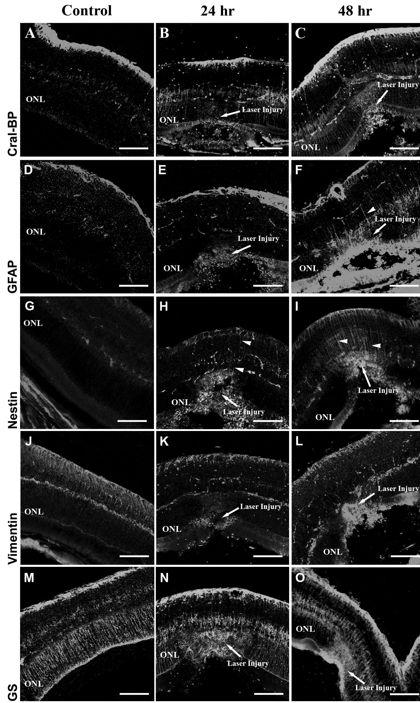
 Figure 1 of
Tackenberg, Mol Vis 2009; 15:1886-1896.
Figure 1 of
Tackenberg, Mol Vis 2009; 15:1886-1896.