Figure 5. Serial electron micrographs
through 4 retracted spherules in 7-day detached retina. Scale bar (in A)
for A-D represents 2 μm. A: Spherules 1 and 2 contain
synaptic ribbons (arrows), each apposing the top of their respective
synaptic invaginations. Only mitochondria are seen in spherules 3 and
4. Arrowheads line the interfaces of apposing Müller cells. Swollen
HCat processes (*) that lack polysomes can be differentiated from
Müller cell processes (M) that have many. B: In the next
section, the opposing lobes at each synaptic ribbon in spherules 1 and
2 are visible. They contain vesicles as do the swollen HCat processes
(*) from which they arise. The hilus of spherule 1 can be seen. Another
HCat process directly apposes spherule 3. C: Two sections later
the openings to the synaptic invaginations of spherules 1 and 2 are
obvious. The hilus of spherule 2 actually faces the outer nuclear layer
instead of the outer plexiform layer. The horizontal cell (HC) lateral
elements innervating spherules 1 and 2 connect with numerous swollen
HCat processes (*) running along apposing Müller cell surfaces.
Synaptic ribbons (arrows) now can be seen in spherules 3 and 4, both of
which directly appose HCat processes. D: Two sections later,
one lobe of the synaptic invagination can be seen in spherule 3 while
spherule 4 makes open contact with its innervating HCat processes (*).
The innervation of spherule 1 by HC lateral elements is still evident,
while that of spherule 2 is not. There is no clear evidence that any of
these 4 spherules have contact with rod bipolar dendrites.
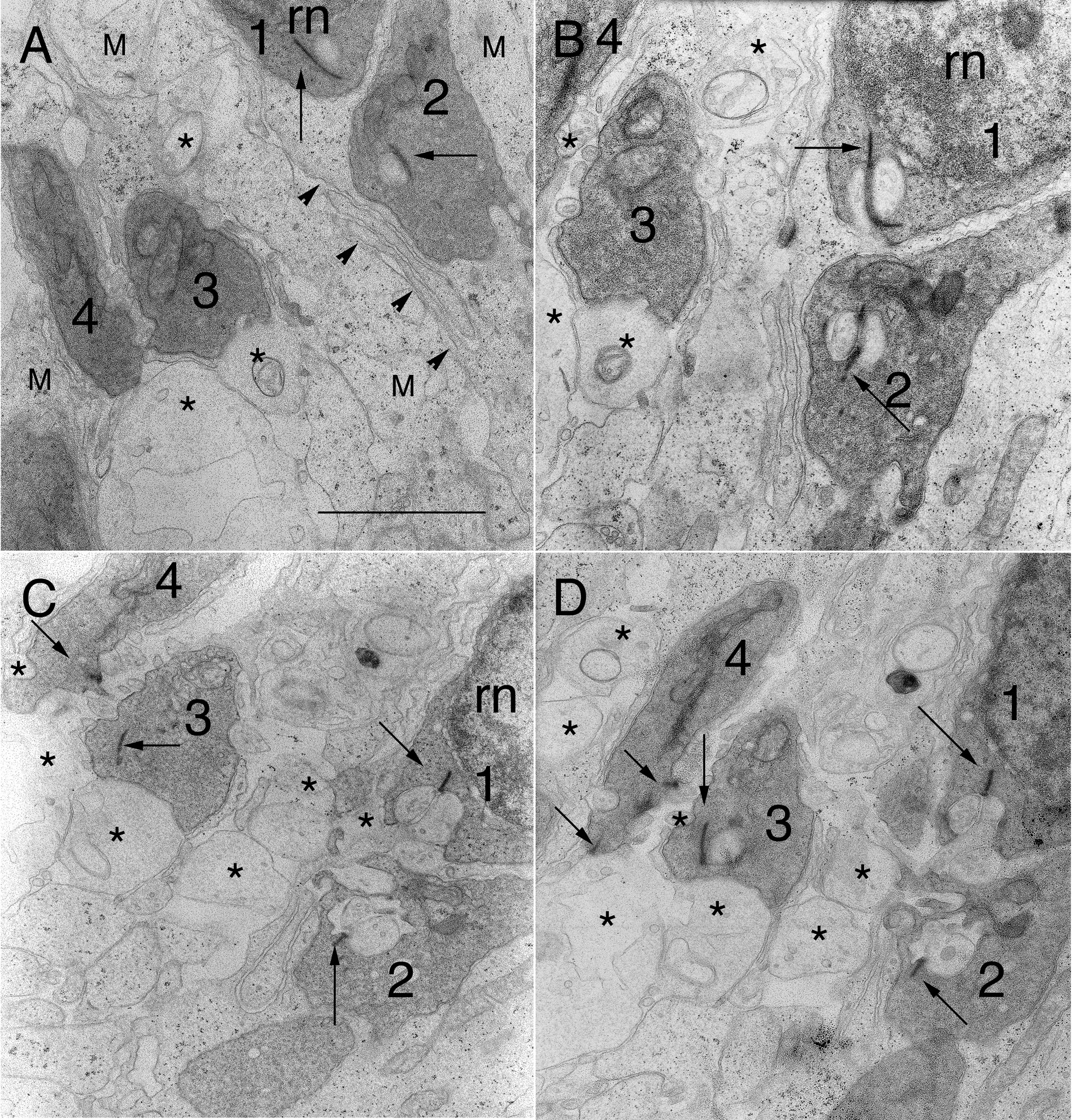
 Figure 5 of Linberg, Mol Vis 2009; 15:10-25.