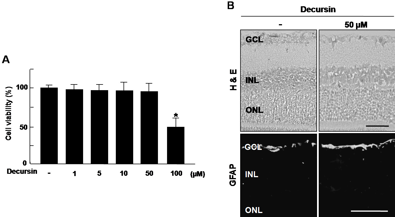
Figure 4. Decursin induced no cytotoxicity
in retinal endothelial cells and no retinal toxicity. A: Human
retinal microvascular endothelial cells were treated with 1–100 µM of
decursin then incubated for 48 h. Cell viability was measured by
3-(4,5-dimethylthiazol-2-yl)-2,5-diphenyltetrazolium bromide (MTT)
assay. Each value represents means±SEM from three independent
experiments. The asterisk indicates a p<0.05. B: We injected
intravitreously 50 µM decursin into mouse eyes, then enucleated the
globes three days after treatment. Hematoxylin & eosin staining and
immunohistochemistry for glial fibrillary acidic protein were
performed. Figures were selected as representative data from three
independent experiments with similar results. Scale bars equal 50 µm.
Abbreviations: ganglion cell layer (GCL), hematoxylin and eosin
(H&E), inner nuclear layer (INL), outer nuclear layer (ONL).

 Figure 4 of Kim, Mol Vis 2009; 15:1868-1875.
  Figure 4 of Kim, Mol Vis 2009; 15:1868-1875.