Figure 3. Decursin inhibits retinal neovascularization in oxygen-induced retinopathy. A, B: Retinal vasculature in control mice and decursin-treated mice with oxygen-induced retinopathy (OIR) was evaluated by fluorescence
angiography using fluorescein-conjugated dextran. Whole-mount retinal preparation from postnatal day 17 (P17) control mice
(A) and mice subjected to OIR and treated with 10 µM decursin (B) was performed after 1 h of perfusion with fluorescein-conjugated dextran. Arrows indicate neovascular tufts of intravitreous
neovascularization. Figures were selected as representative data from three independent experiments with similar results.
Scale bars equal 50 µm. Hematoxylin-stained cross-sections were prepared from P17 control mice (C) and mice subjected to OIR and treated with 10 μM of decursin (D). Arrows indicate the vascular lumens of new vessels growing into the vitreous. Figures were selected as representative data
from three independent experiments with similar results. Scale bars equal 100 µm. E: Each value represents the mean (±SD) of three independent experiments. The asterisk indicates a p<0.05.
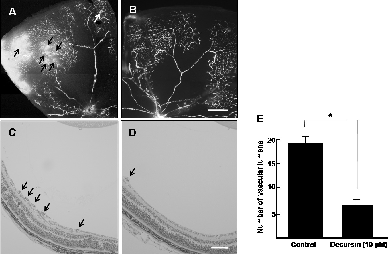
 Figure 3 of
Kim, Mol Vis 2009; 15:1868-1875.
Figure 3 of
Kim, Mol Vis 2009; 15:1868-1875.