Figure 4. Osmotic swelling properties of
Müller cells from A1AR−/− and wildtype (Wt)
mice. The cross-sectional area of Müller cell somata was measured after
a 4 min perfusion of retinal slices with a hypoosmolar solution (in the
absence of barium), and are expressed in percent of the soma size
measured before hypotonic challenge (100%). A: Hypoosmotic
exposure evoked swelling of Müller cell bodies in retinal slices from A1AR−/−
mice. This was observed in freshly isolated retinas and retinal organ
cultures. In contrast, in tissues from wild-type animals, swelling was
induced in retinal organ cultures but not in freshly isolated retinas. B:
Müller cells in retinal slices derived from A1AR−/−
mice displayed a swelling of their cell bodies under hypoosmotic
conditions (control). In slices of these mice, 10 µM adenosine did not
prevent the swelling of the cells. The swelling was diminished in the
presence of a cAMP-enhancing cocktail containing 100 µM pCPT-cAMP, 10
µM forskolin, and 100 µM IBMX. Significant differences versus freshly
isolated cells from wild-type mice (the asterisk indicates a
p<0.001). Significant swelling-inhibitory effect (the filled circle
indicates a p<0.001).
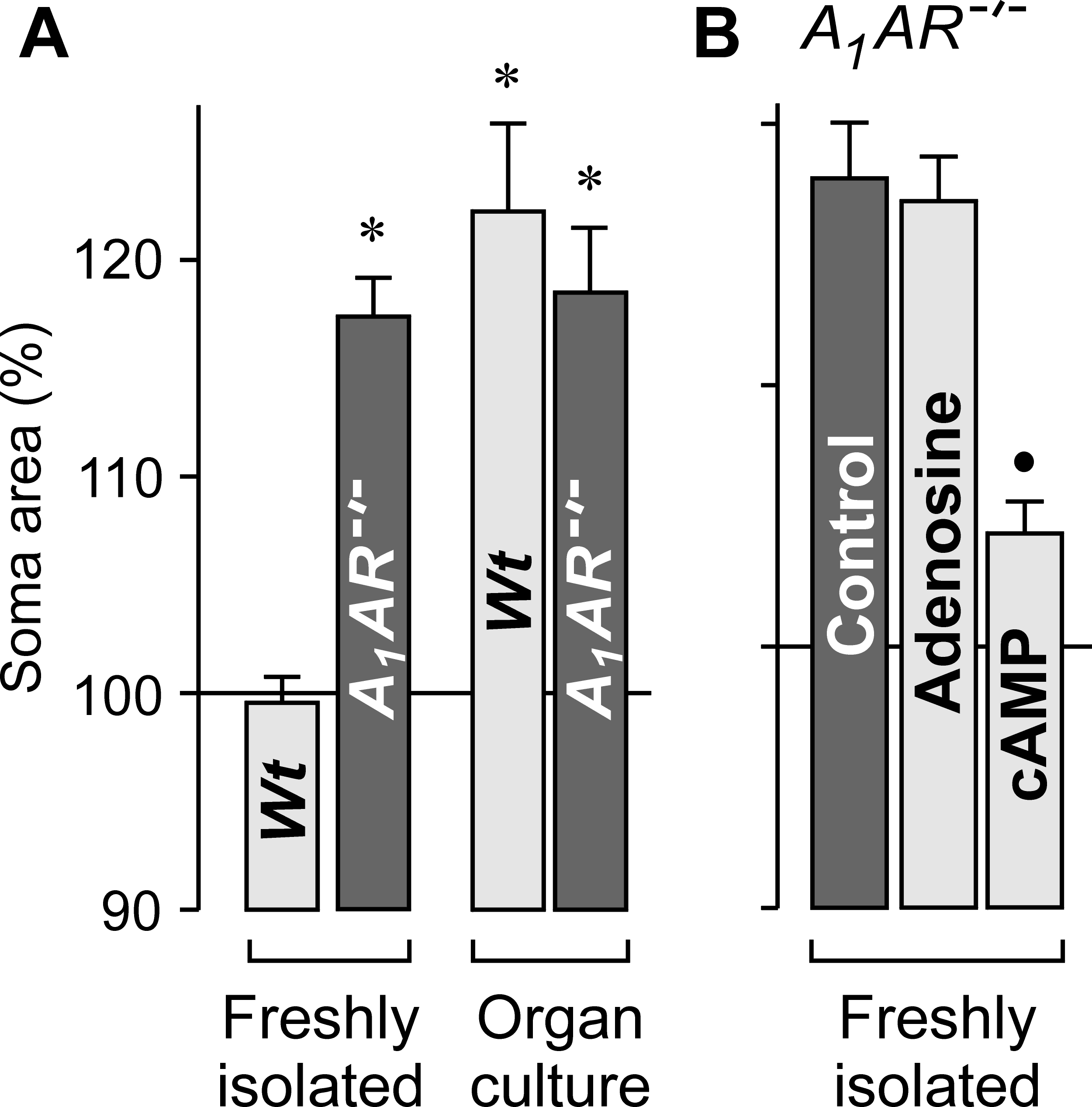
 Figure 4 of Wurm, Mol Vis 2009; 15:1858-1867.