Figure 3. Endogenous adenosine signaling
is required to prevent the osmotic swelling of Müller cells. Data were
obtained in freshly isolated retinal slices (A) and Müller cells
(B) from wild-type mice. Perfusion of the slices or cells with a
hypoosmolar solution containing 1 mM barium chloride, 100 nM DPCPX, a A1
receptor blocker, 100 µM AOPCP, an ecto-5′-nucleotidase inhibitor, 10
µM clofilium, a potassium channel blocker, or 100 µM NPPB, a chloride
channel blocker, resulted in a swelling of Müller cell somata. C:
Original records of a dye-filled isolated Müller cell before (left) and
during (right) perfusion with the barium-containing hypoosmolar
solution. The arrowhead indicates the cell soma, and the arrow refers
to the cell endfoot. The scale bar equals 5 µm. Data are mean (±SEM)
soma areas (n=7–14 cells per bar) which were measured after a 4 min
perfusion of the hypoosmolar solution. Data are expressed in percent of
the soma size measured before hypotonic challenge (100%). Significant
differences versus control (the asterisk indicates a p<0.05, the
double asterisk indicates a p<0.01, and the triple asterisk
indicates a p<0.001).
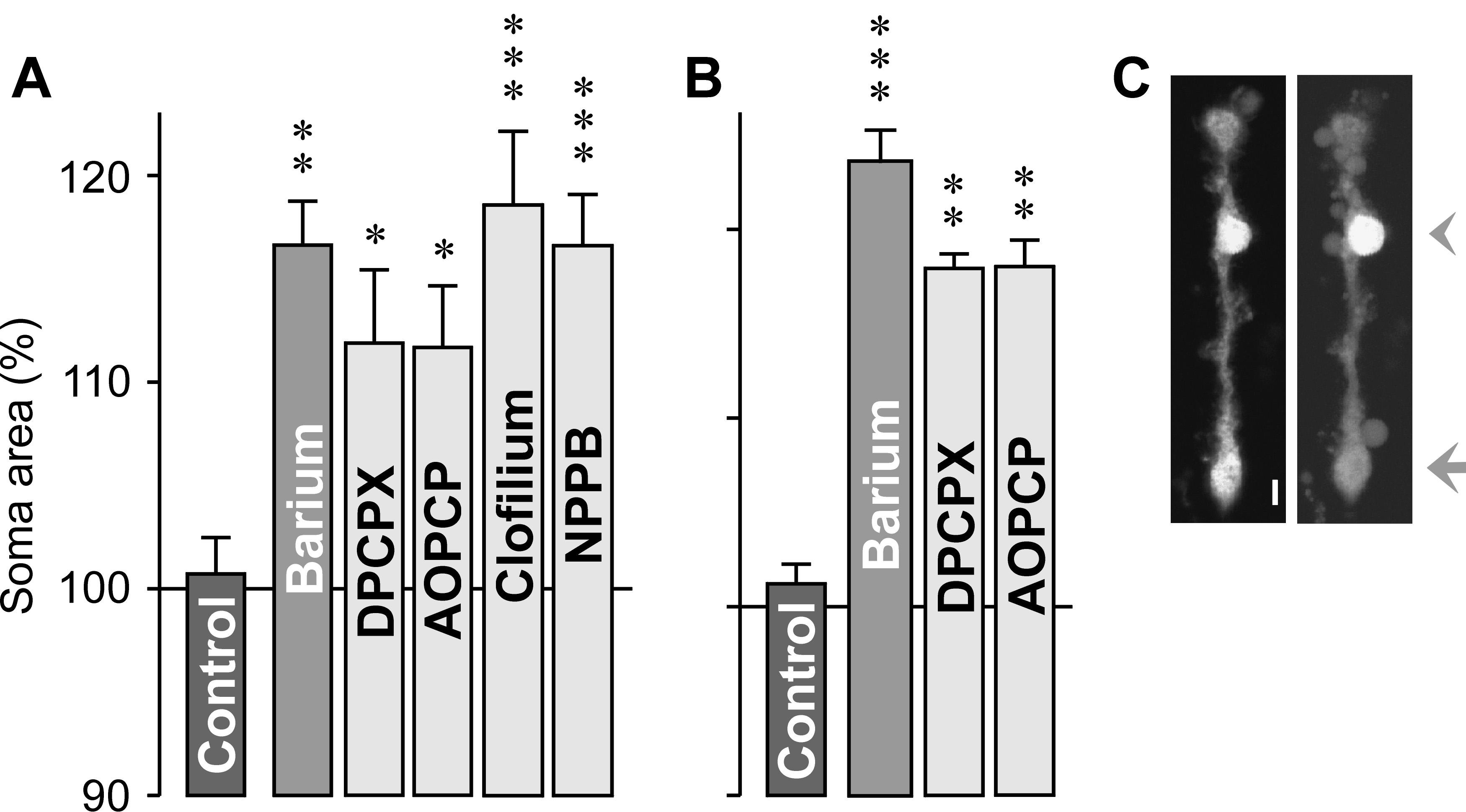
 Figure 3 of Wurm, Mol Vis 2009; 15:1858-1867.