Figure 1. Osmotic swelling of Müller glial
cells in slices of the wild-type mouse retina. A: Perfusion of
slices of a freshly isolated retina with a hypoosmolar solution,
containing 60% of control osmolarity, in the absence (control) and
presence of 1 mM barium chloride resulted in a time-dependent swelling
of Müller cell somata. The diagram displays the mean (±SEM)
cross-sectional area of the somata (n=3 each). Inset images in A
show original records of dye-filled Müller cell somata obtained before
(left) and during (right) perfusion with the barium-containing
hypoosmolar solution. Scale bars represents 5 µm. B:
Hypoosmotic exposure for 4 min evoked swelling of Müller cell somata in
slices of freshly isolated retinas in the presence but not absence of 1
mM barium chloride. Müller cells in slices of retinal organ cultures
displayed soma swelling under both conditions. Data are mean (±SEM)
soma areas (n=7–21 cells per bar) expressed in percent of the soma size
recorded before hypoosmotic challenge (100%). Significant increases in
the soma area: The double asterisk indicates a p<0.01 and the triple
asterisk indicates a p<0.001.
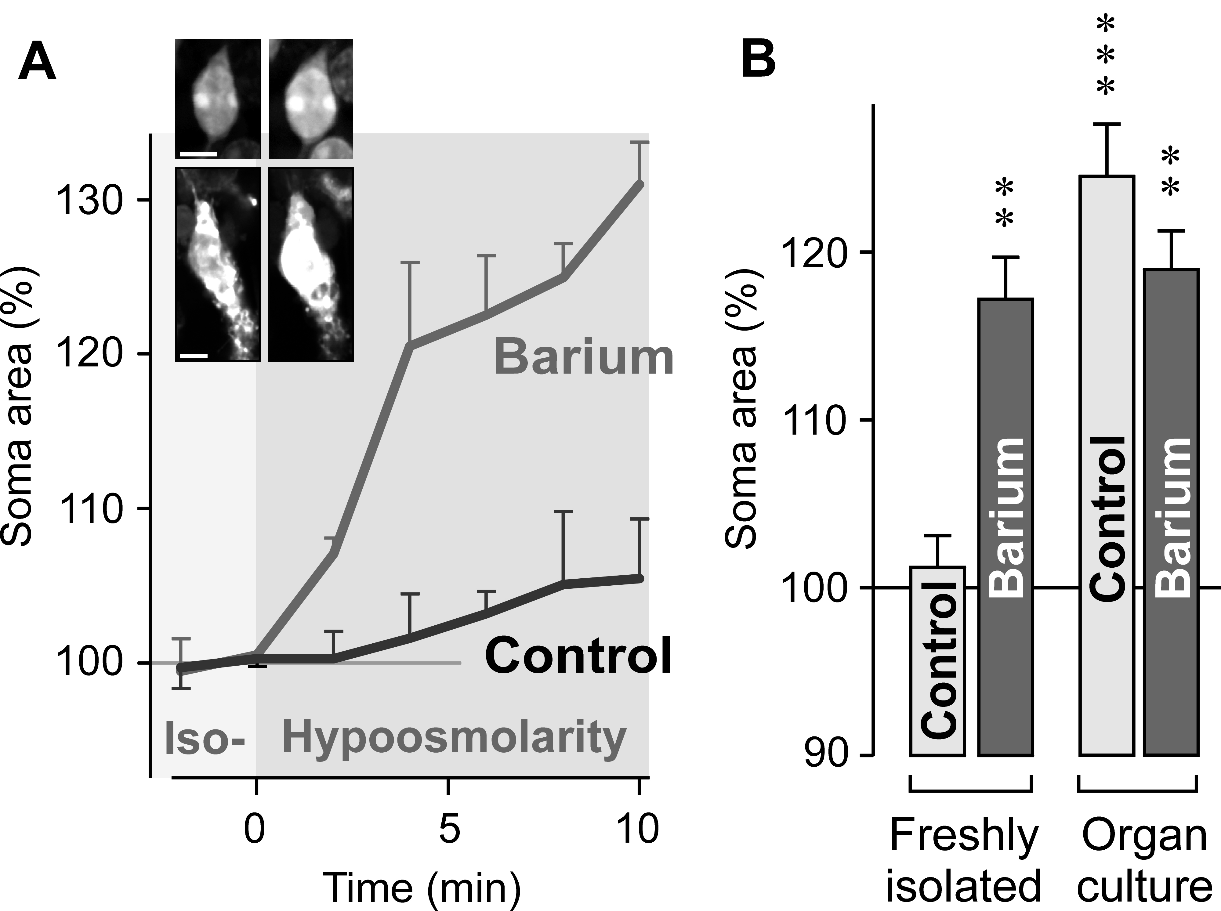
 Figure 1 of Wurm, Mol Vis 2009; 15:1858-1867.