Figure 8. Primate and mouse CDH23 protein
in inner ear and retina. A-C: western blot analyses of proteins
separated on 3%–8% tris-acetate gels using the cytoplasmic domain
antibody, TF7. A: Western analysis, using protein extracts from
P0, and P90 wild-type and mutant mouse inner ear and retina. An
approximately 350 kDa band corresponding to the largest CDH23
protein isoform was only detected in P0 mouse inner ear (arrow head).
Faster migrating protein bands were present in wild-type and Cdh23v-6J
and may represent lower molecular weight CDH23 protein isoforms (such
as CDH23_V3). Tissue specific variation of these isoforms is better
visualized in the western blot shown in panel B. B: Western
analysis, using protein extracts from P4, and P60 wild-type mouse inner
ear and retina. The high molecular weight band at roughly 350 kDa
(arrowhead) was detected in the P4 inner ear protein sample. Traces of
this band were also detected in the P60 inner ear protein sample. The
faster migrating bands detected show variability in their appearance
between young and adult tissue as well as variability between the inner
ear and retina. C: Western analysis, using protein extracts
from P0 mouse retina, human retina, and monkey retina. The high
molecular weight band (arrow head A-C) detected in P0 wild-type
mouse inner ear corresponds in size to the largest band detected in
human
and monkey retinas (arrowhead). β-actin was used as a loading control;
size standards are given in kDa.
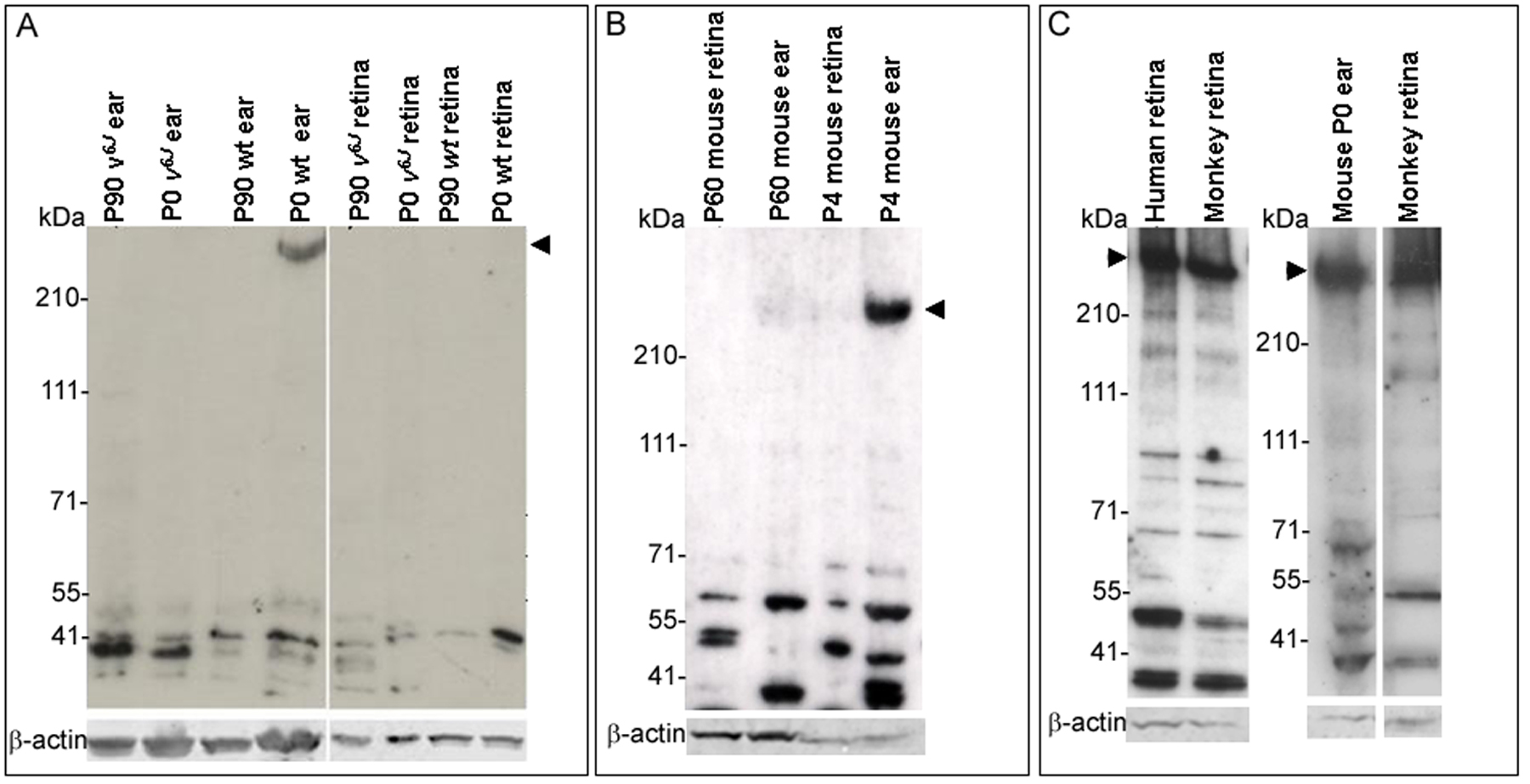
 Figure 8 of Lagziel, Mol Vis 2009; 15:1843-1857.