Figure 7. Immunolocalization of CDH23 in
photoreceptor cells of monkey retina. A: CDH23 immunoreactivity
was detected with antisera TF7 in the IS and in the synaptic terminals
of the OPL of photoreceptor cells. Some staining is evident in the
photoreceptor cell bodies of the ONL and INL. B: No
immunoreactivity was observed in the controls stained only with
secondary antibody. C: In a higher magnification, CDH23
staining is visible in the evenly spaced IS of cone-shaped
photoreceptors (asterisks) and in the regions between the IS and OS
where the ciliary apparatus is present (arrows). Rod photoreceptors are
also positively stained (seen as thin long strings). The dark brown
pigment in the upper portion of A and B is melanin of
the RPE. C: Scale bars represent 5 µM. Magnification: A, B
400× and C 1000×. Abbreviations: retinal pigment epithelium
(RPE), outer segment (OS), inner segment (IS), outer nuclear layer
(ONL), outer plexiform layer (OPL), inner nuclear layer (INL).
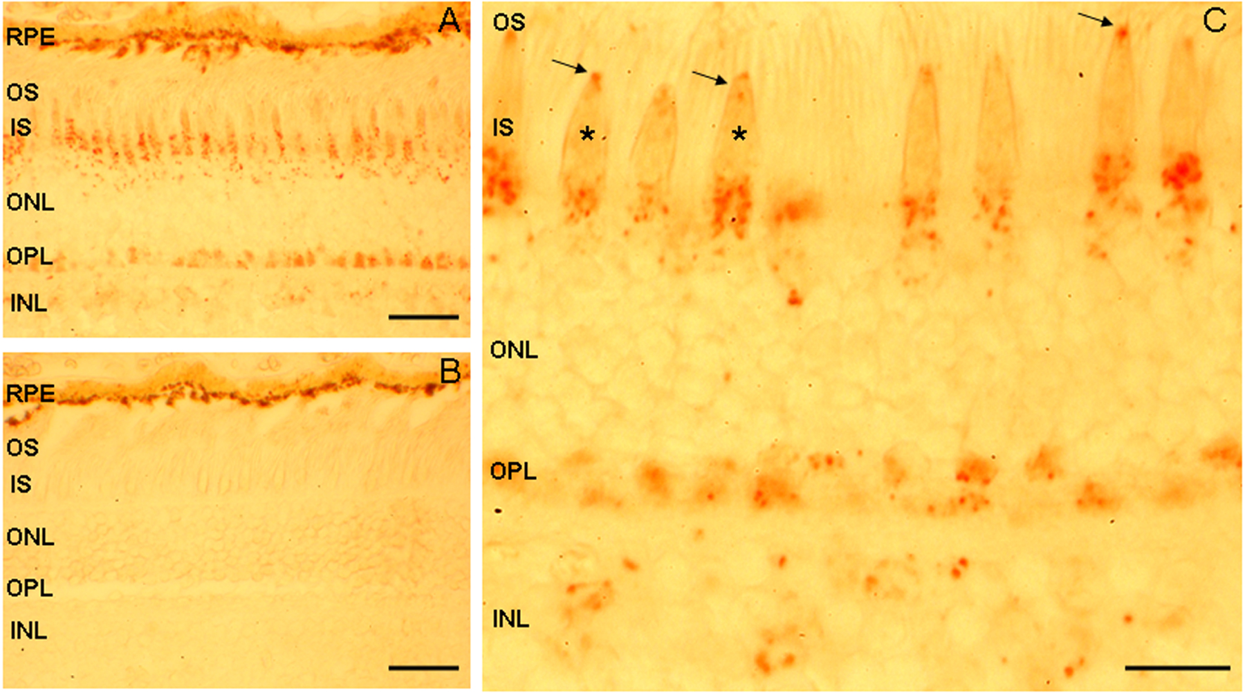
 Figure 7 of Lagziel, Mol Vis 2009; 15:1843-1857.