Figure 6. CDH23 localizes with SNAP25 to
the synaptic region of mouse auditory and vestibular hair cells. In A-I
nuclei were stained with DAPI (blue). SNAP25 is shown in red, and CDH23
in green. A: The synaptic region of utricular hair cells
stained with SNAP25. B: CDH23_V3 detected with antisera TF648. C:
In the merge image (C) of panels A and B,
CDH23_V3 shows partial colocalization with SNAP25 in the synaptic
region of utricular hair cells. The insets in A-C show a
twofold magnification of a hair cell (asterisk) synaptic area. In the
merge image (F) of panels D and E, CDH23_3 and
SNAP25 show partial colocalization in the synaptic region of inner
(IHC) and outer (OHC) hair cells. G: SNAP25 staining of IHC and
OHC synaptic fibers. H: CDH23 is detected with antiserum TF7 in
the synaptic fibers of IHC and OHC. I: In the merge image (I)
of panels G and H, CDH23 shows partial colocalization
with SNAP25 in the region of the synaptic fibers of IHC and OHC. In G-I,
the insets are a twofold magnification of the region (asterisk), where
CDH23 localizes in the synaptic fibers. Scale bars equal 5 µm.
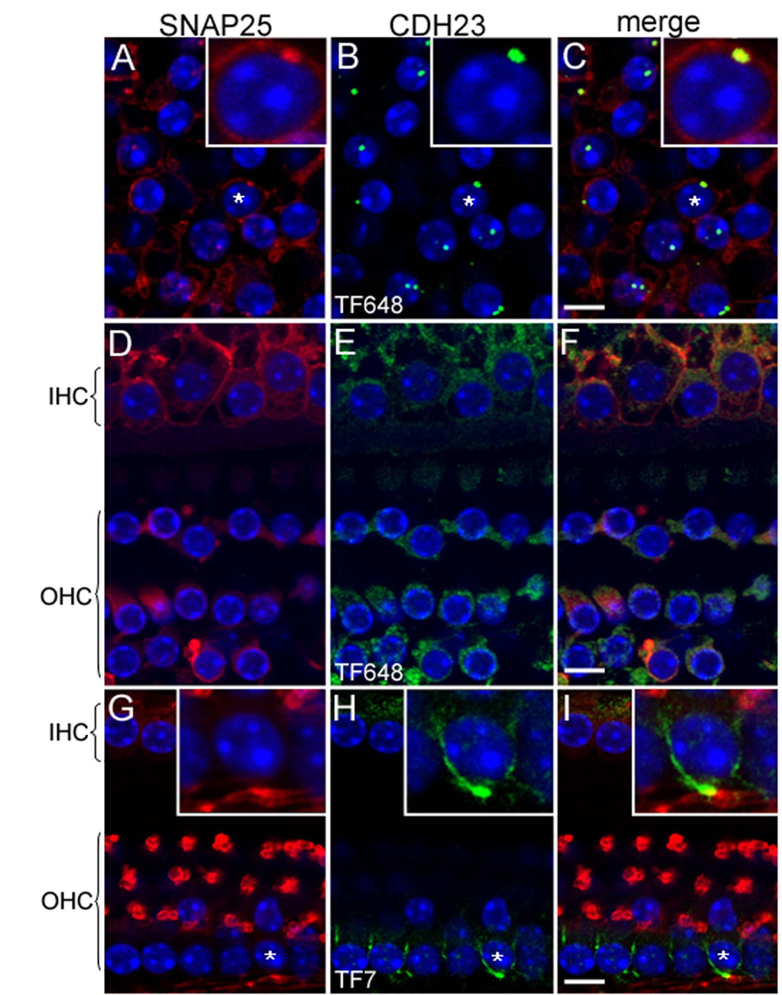
 Figure 6 of Lagziel, Mol Vis 2009; 15:1843-1857.