Figure 5. Immunoelectron microscopic localization of CDH23 in mouse rod photoreceptor cells. Electron micrographs show CDH23 labeling
with TF7 antibody in ultrathin sections through parts of mouse photoreceptor cells. A-C: CDH23 labeling is detected in the basal body (BB) and centriole (CE) of the apical inner segment of photoreceptor cells.
A, C: The connecting cilium (CC) is not labeled. Scale bar represents 0.2 µm.
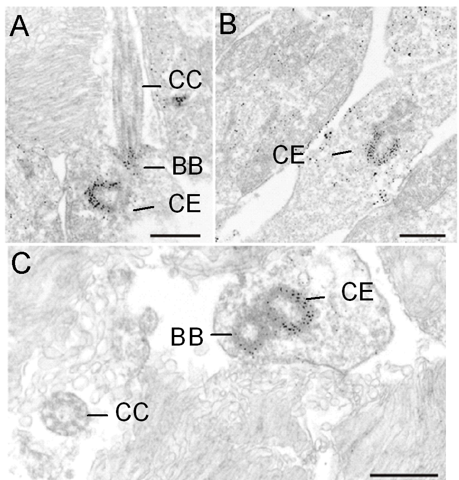
 Figure 5 of
Lagziel, Mol Vis 2009; 15:1843-1857.
Figure 5 of
Lagziel, Mol Vis 2009; 15:1843-1857.