Figure 3. Different CDH23 protein isoforms
localize to distinct cell layers of the retina. A, E, I, and M
are differential interference contrast (DIC) images of longitudinal
cryosections (B-D, F-H, J-L and N-P, respectively)
through adult mouse retina showing photoreceptors cell layers. Indirect
immunofluorescence labeling of CDH23 with the cytoplasmic domain
antibody TF7 (B) revealed CDH23 in the ciliary region of
photoreceptor cells, the ONL, and in the OPL. Double labeling with
antibodies TF7 and centrin, a ciliary marker (C), revealed
colocalization of CDH23 and centrin in the ciliary apparatus of the
photoreceptor cells (D). F-H: Double labeling with
antisera TF7 (F) and the synaptic protein PSD-95 with antisera
PSD95 (G) revealed CDH23 colocalization with PSD-95 in the
synaptic terminals in the OPL of photoreceptors cells (H).
Double labeling with CDH23_V3 specific antibodies TF648 and TF649 (J,
N) and PSD95 (K, O) revealed that CDH23_V3 was detected only
in the OPL where it colocalized with PSD-95 (L, P). Scale bars
represent 10 µm. Abbreviations: retinal pigment epithelium (RPE), outer
segment (OS), inner segment (IS), outer nuclear layer (ONL), outer
plexiform layer (OPL), and inner nuclear layer (INL).
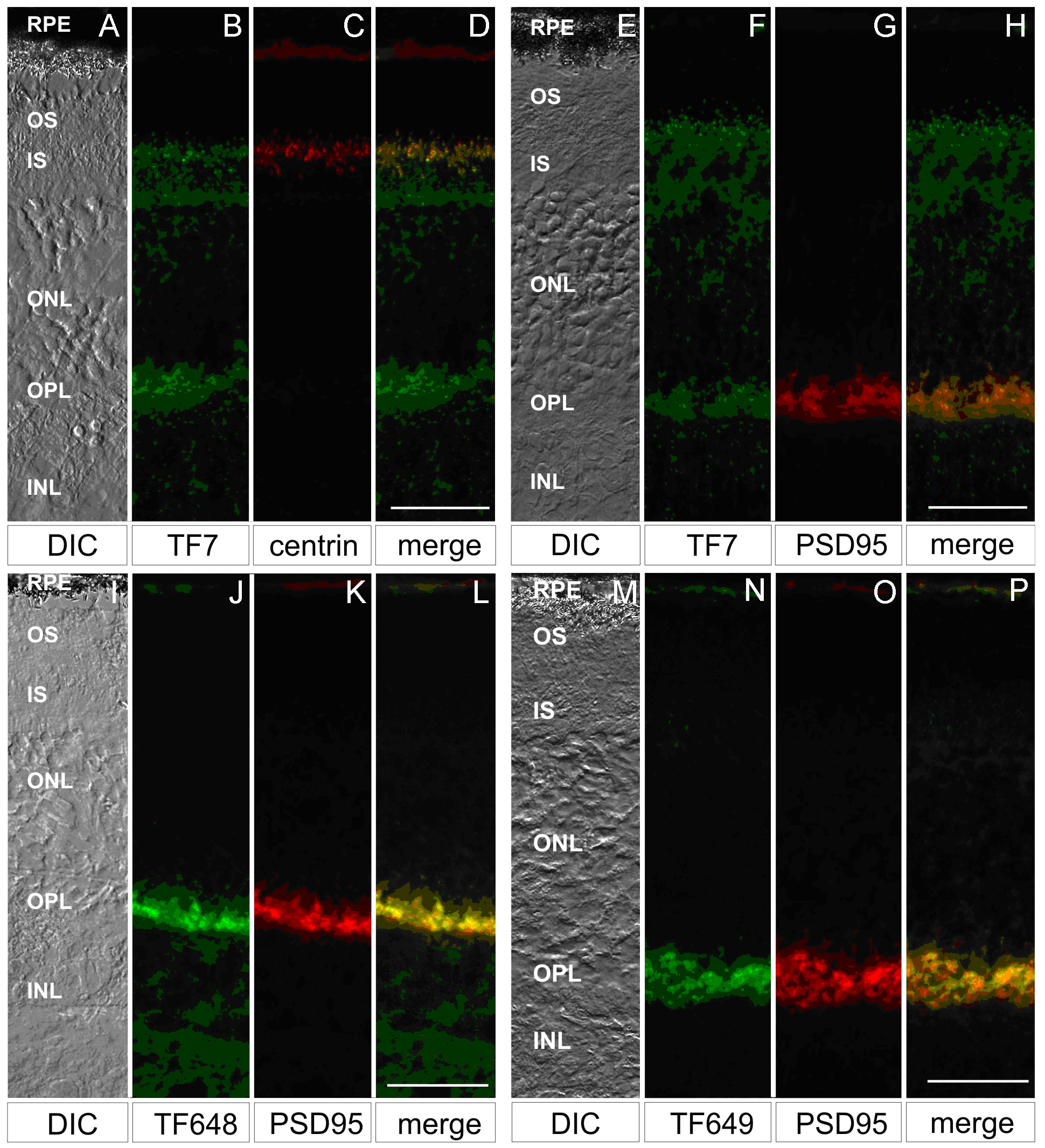
 Figure 3 of Lagziel, Mol Vis 2009; 15:1843-1857.