Figure 2. Immunohistochemical
investigation of retinal cell type expressing GFP. A to C
are conventional fluorescent microscopy images and D to F
are confocal microscopy images. A and B: Sections
through the ssAAV2/5 injected eye from the peripheral retina away for
the injection site (A) and through the injection site (B).
C: A section through the injection site in a scAAV2/5 injected
eye. D: A section through the injected region of a scAAV2/5
eye. The Z stack is shown on the right. Co-localization of hCAR
immunoreactivity and GFP expression is present in some, but not all of
the cone photoreceptors. Arrows indicate two hCAR positive cones that
do not have observable endogenous GFP expression. E: The rod
bipolar cells (which are PKC immunoreactive) are not expressing GFP. F:
There is co-localization of GFP expression and PSD95 immunoreactivity
in photoreceptor terminals. Abbreviations: ONL, outer nuclear layer ;
INL, inner nuclear layer; GCL, ganglion cell layer, PKC, protein kinase
C (rod bipolar cell marker); hCAR, human cone arrestin (a cone marker);
GFP, green fluorescent protein; PSD95, post synaptic density protein 95
(a marker for rod and cone spherules). Scale bar equals 50 μm.
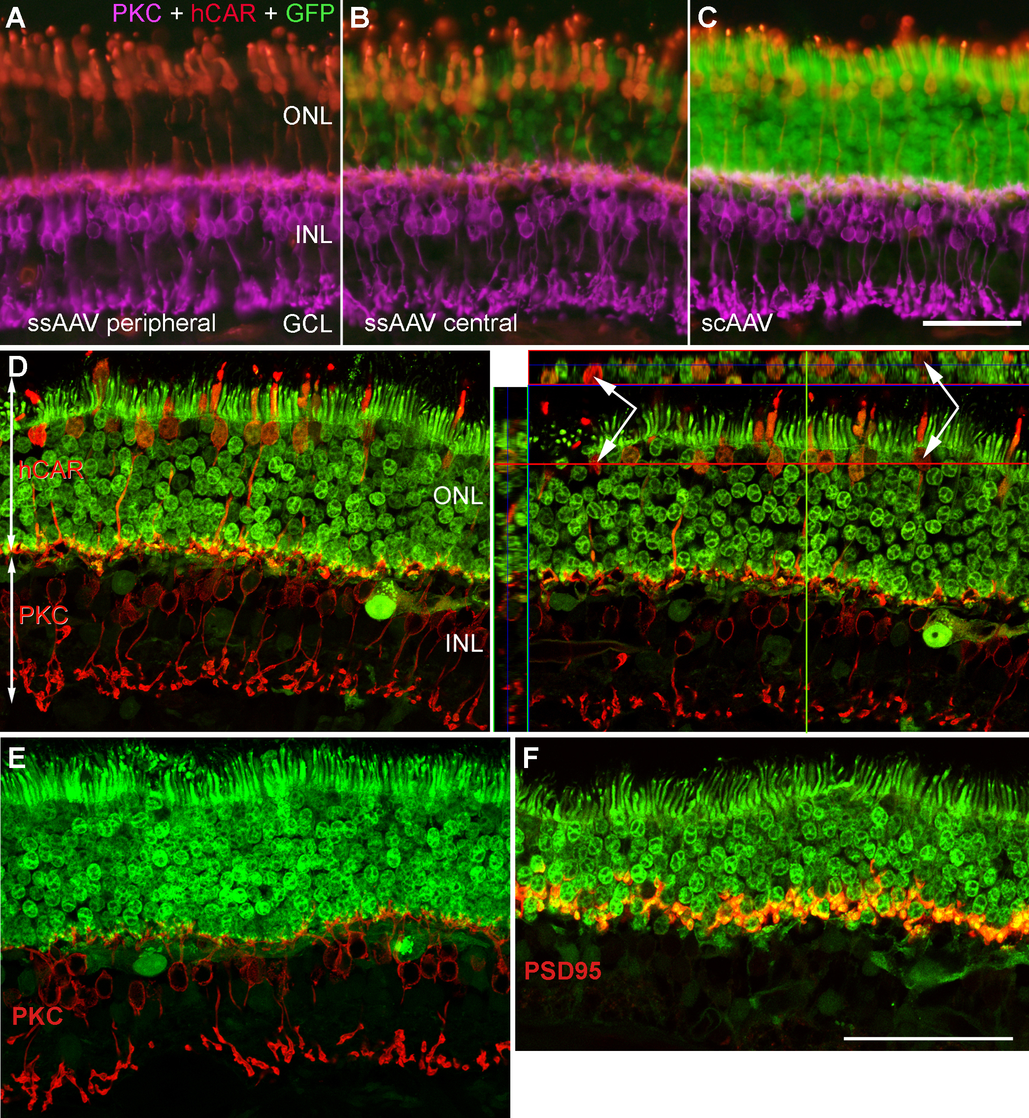
 Figure 2 of Petersen-Jones, Mol Vis 2009; 15:1835-1842.