Figure 1. Comparison of GFP expression in
scAAV2/5 and ssAAV2/5 injected eyes (dog 1). A: Pre-INJ shows
the fundus appearance before subretinal injection. post-INJ images were
taken immediately after subretinal injection and show the resulting
retinal detachment. The images day 6 to day 54 were taken using
identical fluorescein angiography settings. The lower images (day 6 and
11 for scAAV2/5 and day 6 to 54 for ssAAV2/5) were adjusted for
brightness and contrast to the same degree. GFP expression in the
scAAV2/5 injected eye was detectable from day 6 whereas in the ssAAV2/5
injected eye it was not apparent until day 32. GFP expression was
stronger in the scAAV2/5 injected eye. B: Comparison of GFP
fluorescence in retinal cross sections from the center of the injected
regions of both eyes of dog 1 (obtained with identical microscope and
camera settings). Scale bar equals 50 µm.
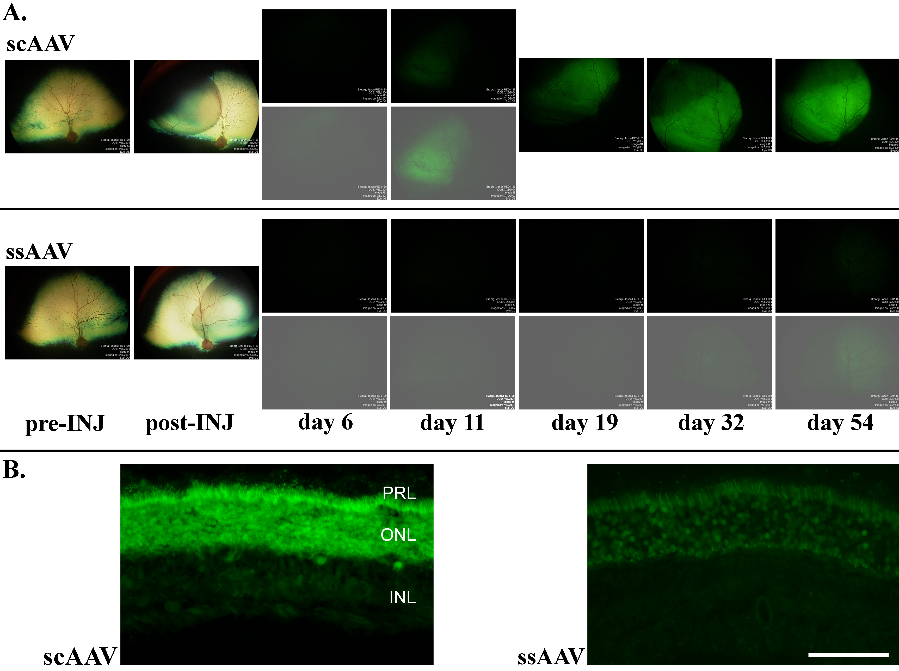
 Figure 1 of Petersen-Jones, Mol Vis 2009; 15:1835-1842.